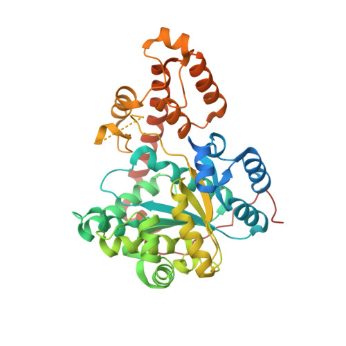
2IP1 | pdb_00002ip1

Crystal Structure Analysis of S. cerevisiae Tryptophanyl tRNA Synthetase


Experimental Data Snapshot

  • Method: X-RAY DIFFRACTION
  • Resolution: 1.80 Å
  • R-Value Free: 
    0.214 (Depositor), 0.216 (DCC) 
  • R-Value Work: 
    0.181 (Depositor), 0.181 (DCC) 
  • R-Value Observed: 
    0.183 (Depositor) 

wwPDB Validation 3D Report Full Report

Validation slider image for 2IP1

This is version 1.4 of the entry. See complete history. 

Literature

Blocking S-adenosylmethionine synthesis in yeast allows selenomethionine incorporation and multiwavelength anomalous dispersion phasing.

Malkowski, M.G., Quartley, E., Friedman, A.E., Babulski, J., Kon, Y., Wolfley, J., Said, M., Luft, J.R., Phizicky, E.M., DeTitta, G.T., Grayhack, E.J.

(2007) Proc Natl Acad Sci U S A 104: 6678-6683

  • DOI: https://doi.org/10.1073/pnas.0610337104
  • Primary Citation Related Structures: 
    2IP1

  • PubMed Abstract: 

    Saccharomyces cerevisiae is an ideal host from which to obtain high levels of posttranslationally modified eukaryotic proteins for x-ray crystallography. However, extensive replacement of methionine by selenomethionine for anomalous dispersion phasing has proven intractable in yeast. We report a general method to incorporate selenomethionine into proteins expressed in yeast based on manipulation of the appropriate metabolic pathways. sam1(-) sam2(-) mutants, in which the conversion of methionine to S-adenosylmethionine is blocked, exhibit reduced selenomethionine toxicity compared with wild-type yeast, increased production of protein during growth in selenomethionine, and efficient replacement of methionine by selenomethionine, based on quantitative mass spectrometry and x-ray crystallography. The structure of yeast tryptophanyl-tRNA synthetase was solved to 1.8 A by using multiwavelength anomalous dispersion phasing with protein that was expressed and purified from the sam1(-) sam2(-) strain grown in selenomethionine. Six of eight selenium residues were located in the structure.


  • Organizational Affiliation: 
    • Center for Pediatric Biomedical Research and Departments of Environmental Medicine and Biochemistry and Biophysics, University of Rochester Medical School, Rochester, NY 14642, USA.

Macromolecule Content 

  • Total Structure Weight: 50.2 kDa 
  • Atom Count: 3,386 
  • Modeled Residue Count: 371 
  • Deposited Residue Count: 432 
  • Unique protein chains: 1

Macromolecules

Find similar proteins by:|  3D Structure
Entity ID: 1
MoleculeChains  Sequence LengthOrganismDetailsImage
Tryptophanyl-tRNA synthetase432Saccharomyces cerevisiaeMutation(s): 0 
Gene Names: WRS1
EC: 6.1.1.2
UniProt
Find proteins for Q12109 (Saccharomyces cerevisiae (strain ATCC 204508 / S288c))
Explore Q12109 
Go to UniProtKB:  Q12109
Entity Groups
Sequence Clusters30% Identity50% Identity70% Identity90% Identity95% Identity100% Identity
UniProt GroupQ12109
Sequence Annotations
Expand
Reference Sequence

Experimental Data & Validation

Experimental Data

  • Method: X-RAY DIFFRACTION
  • Resolution: 1.80 Å
  • R-Value Free:  0.214 (Depositor), 0.216 (DCC) 
  • R-Value Work:  0.181 (Depositor), 0.181 (DCC) 
  • R-Value Observed: 0.183 (Depositor) 
Space Group: P 43 21 2
Unit Cell:
Length ( Å )Angle ( ˚ )
a = 56.35α = 90
b = 56.35β = 90
c = 313.64γ = 90
Software Package:
Software NamePurpose
REFMACrefinement
Blu-Icedata collection
MOSFLMdata reduction
SCALAdata scaling
SnBphasing

Structure Validation

View Full Validation Report


Entry History 

Deposition Data

Revision History  (Full details and data files)

  • Version 1.0: 2007-06-26
    Type: Initial release
  • Version 1.1: 2008-05-01
    Changes: Version format compliance
  • Version 1.2: 2011-07-13
    Changes: Advisory, Version format compliance
  • Version 1.3: 2017-10-18
    Changes: Refinement description
  • Version 1.4: 2024-02-21
    Changes: Data collection, Database references, Derived calculations