2IMG

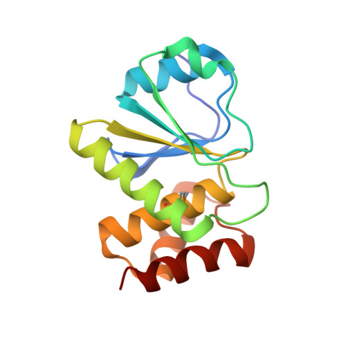
Crystal structure of dual specificity protein phosphatase 23 from Homo sapiens in complex with ligand malate ion


Experimental Data Snapshot

  • Method: X-RAY DIFFRACTION
  • Resolution: 1.93 Å
  • R-Value Free: 0.229 
  • R-Value Work: 0.195 
  • R-Value Observed: 0.195 

wwPDB Validation   3D Report Full Report


This is version 1.4 of the entry. See complete history


Literature

Structure of human dual specificity protein phosphatase 23, VHZ, enzyme-substrate/product complex.

Agarwal, R.Burley, S.K.Swaminathan, S.

(2008) J Biol Chem 283: 8946-8953

  • DOI: https://doi.org/10.1074/jbc.M708945200
  • Primary Citation of Related Structures:  
    2IMG

  • PubMed Abstract: 

    Protein phosphorylation plays a crucial role in mitogenic signal transduction and regulation of cell growth and differentiation. Dual specificity protein phosphatase 23 (DUSP23) or VHZ mediates dephosphorylation of phospho-tyrosyl (pTyr) and phospho-seryl/threonyl (pSer/pThr) residues in specific proteins. In vitro, it can dephosphorylate p44ERK1 but not p54SAPK-beta and enhance activation of c-Jun N-terminal kinase (JNK) and p38. Human VHZ, the smallest of the catalytically active protein-tyrosine phosphatases (PTP) reported to date (150 residues), is a class I Cys-based PTP and bears the distinctive active site signature motif HCXXGXXRS(T). We present the crystal structure of VHZ determined at 1.93A resolution. The polypeptide chain adopts the typical alphabetaalpha PTP fold, giving rise to a shallow active site cleft that supports dual phosphorylated substrate specificity. Within our crystals, the Thr-135-Tyr-136 from a symmetry-related molecule bind in the active site with a malate ion, where they mimic the phosphorylated TY motif of the MAPK activation loop in an enzyme-substrate/product complex. Analyses of intermolecular interactions between the enzyme and this pseudo substrate/product along with functional analysis of Phe-66, Leu-97, and Phe-99 residues provide insights into the mechanism of substrate binding and catalysis in VHZ.


  • Organizational Affiliation

    Biology Department, Brookhaven National Laboratory, Upton, NY 11973, USA.


Macromolecules
Find similar proteins by:  (by identity cutoff)  |  3D Structure
Entity ID: 1
MoleculeChains Sequence LengthOrganismDetailsImage
Dual specificity protein phosphatase 23151Homo sapiensMutation(s): 1 
Gene Names: DUSP23
EC: 3.1.3.48 (PDB Primary Data), 3.1.3.16 (PDB Primary Data)
UniProt & NIH Common Fund Data Resources
Find proteins for Q9BVJ7 (Homo sapiens)
Explore Q9BVJ7 
Go to UniProtKB:  Q9BVJ7
PHAROS:  Q9BVJ7
GTEx:  ENSG00000158716 
Entity Groups  
Sequence Clusters30% Identity50% Identity70% Identity90% Identity95% Identity100% Identity
UniProt GroupQ9BVJ7
Sequence Annotations
Expand
  • Reference Sequence
Small Molecules
Ligands 1 Unique
IDChains Name / Formula / InChI Key2D Diagram3D Interactions
MLT
Query on MLT

Download Ideal Coordinates CCD File 
B [auth A]D-MALATE
C4 H6 O5
BJEPYKJPYRNKOW-UWTATZPHSA-N
Modified Residues  1 Unique
IDChains TypeFormula2D DiagramParent
MSE
Query on MSE
A
L-PEPTIDE LINKINGC5 H11 N O2 SeMET
Experimental Data & Validation

Experimental Data

  • Method: X-RAY DIFFRACTION
  • Resolution: 1.93 Å
  • R-Value Free: 0.229 
  • R-Value Work: 0.195 
  • R-Value Observed: 0.195 
  • Space Group: P 21 21 21
Unit Cell:
Length ( Å )Angle ( ˚ )
a = 35.97α = 90
b = 59.253β = 90
c = 64.4γ = 90
Software Package:
Software NamePurpose
CNSrefinement
CBASSdata collection
HKL-2000data reduction
HKL-2000data scaling
SOLVEphasing
SHARPphasing

Structure Validation

View Full Validation Report


Entry History 

Deposition Data

Revision History  (Full details and data files)

  • Version 1.0: 2006-10-17
    Type: Initial release
  • Version 1.1: 2008-03-20
    Changes: Version format compliance
  • Version 1.2: 2011-07-13
    Changes: Version format compliance
  • Version 1.3: 2021-02-03
    Changes: Database references, Derived calculations, Structure summary
  • Version 1.4: 2024-10-30
    Changes: Data collection, Database references, Structure summary