2IIZ | pdb_00002iiz

Crystal structure of putative melanin biosynthesis protein TyrA with bound heme (NP_716371.1) from Shewanella Oneidensis at 2.30 A resolution


Experimental Data Snapshot

  • Method: X-RAY DIFFRACTION
  • Resolution: 2.30 Å
  • R-Value Free: 
    0.266 (Depositor), 0.269 (DCC) 
  • R-Value Work: 
    0.205 (Depositor), 0.210 (DCC) 
  • R-Value Observed: 
    0.208 (Depositor) 

Starting Model: experimental
View more details

wwPDB Validation 3D Report Full Report

Validation slider image for 2IIZ

Ligand Structure Quality Assessment 


This is version 1.7 of the entry. See complete history

Literature

Identification and structural characterization of heme binding in a novel dye-decolorizing peroxidase, TyrA.

Zubieta, C.Joseph, R.Krishna, S.S.McMullan, D.Kapoor, M.Axelrod, H.L.Miller, M.D.Abdubek, P.Acosta, C.Astakhova, T.Carlton, D.Chiu, H.J.Clayton, T.Deller, M.C.Duan, L.Elias, Y.Elsliger, M.A.Feuerhelm, J.Grzechnik, S.K.Hale, J.Han, G.W.Jaroszewski, L.Jin, K.K.Klock, H.E.Knuth, M.W.Kozbial, P.Kumar, A.Marciano, D.Morse, A.T.Murphy, K.D.Nigoghossian, E.Okach, L.Oommachen, S.Reyes, R.Rife, C.L.Schimmel, P.Trout, C.V.van den Bedem, H.Weekes, D.White, A.Xu, Q.Hodgson, K.O.Wooley, J.Deacon, A.M.Godzik, A.Lesley, S.A.Wilson, I.A.

(2007) Proteins 69: 234-243

  • DOI: https://doi.org/10.1002/prot.21673
  • Primary Citation Related Structures: 
    2IIZ

  • PubMed Abstract: 

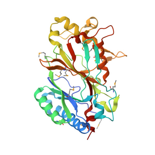
    TyrA is a member of the dye-decolorizing peroxidase (DyP) family, a new family of heme-dependent peroxidase recently identified in fungi and bacteria. Here, we report the crystal structure of TyrA in complex with iron protoporphyrin (IX) at 2.3 A. TyrA is a dimer, with each monomer exhibiting a two-domain, alpha/beta ferredoxin-like fold. Both domains contribute to the heme-binding site. Co-crystallization in the presence of an excess of iron protoporphyrin (IX) chloride allowed for the unambiguous location of the active site and the specific residues involved in heme binding. The structure reveals a Fe-His-Asp triad essential for heme positioning, as well as a novel conformation of one of the heme propionate moieties compared to plant peroxidases. Structural comparison to the canonical DyP family member, DyP from Thanatephorus cucumeris (Dec 1), demonstrates conservation of this novel heme conformation, as well as residues important for heme binding. Structural comparisons with representative members from all classes of the plant, bacterial, and fungal peroxidase superfamily demonstrate that TyrA, and by extension the DyP family, adopts a fold different from all other structurally characterized heme peroxidases. We propose that a new superfamily be added to the peroxidase classification scheme to encompass the DyP family of heme peroxidases.


  • Organizational Affiliation
    • Stanford Synchrotron Radiation Laboratory, Stanford University, Menlo Park, California, USA.

Macromolecule Content 

  • Total Structure Weight: 37.29 kDa 
  • Atom Count: 2,614 
  • Modeled Residue Count: 306 
  • Deposited Residue Count: 312 
  • Unique protein chains: 1

Macromolecules

Find similar proteins by:|  3D Structure
Entity ID: 1
MoleculeChains  Sequence LengthOrganismDetailsImage
Melanin biosynthesis protein TyrA, putative312Shewanella oneidensisMutation(s): 13 
Gene Names: NP_716371.1
UniProt
Find proteins for Q8EIU4 (Shewanella oneidensis (strain ATCC 700550 / JCM 31522 / CIP 106686 / LMG 19005 / NCIMB 14063 / MR-1))
Explore Q8EIU4 
Go to UniProtKB:  Q8EIU4
Entity Groups
Sequence Clusters30% Identity50% Identity70% Identity90% Identity95% Identity100% Identity
UniProt GroupQ8EIU4
Sequence Annotations
Expand
Reference Sequence

Small Molecules

Modified Residues  1 Unique
IDChains TypeFormula2D DiagramParent
MSE
Query on MSE
A
L-PEPTIDE LINKINGC5 H11 N O2 SeMET

Experimental Data & Validation

Experimental Data

  • Method: X-RAY DIFFRACTION
  • Resolution: 2.30 Å
  • R-Value Free:  0.266 (Depositor), 0.269 (DCC) 
  • R-Value Work:  0.205 (Depositor), 0.210 (DCC) 
  • R-Value Observed: 0.208 (Depositor) 
Space Group: P 43 21 2
Unit Cell:
Length ( Å )Angle ( ˚ )
a = 94.837α = 90
b = 94.837β = 90
c = 116.666γ = 90
Software Package:
Software NamePurpose
XSCALEdata scaling
PHASERphasing
REFMACrefinement
MolProbitymodel building
PDB_EXTRACTdata extraction
XDSdata reduction

Structure Validation

View Full Validation Report


Ligand Structure Quality Assessment 


Entry History 

Deposition Data

Revision History  (Full details and data files)

  • Version 1.0: 2006-11-14
    Type: Initial release
  • Version 1.1: 2008-05-01
    Changes: Version format compliance
  • Version 1.2: 2011-07-13
    Changes: Advisory, Version format compliance
  • Version 1.3: 2017-10-18
    Changes: Refinement description
  • Version 1.4: 2023-01-25
    Changes: Database references, Derived calculations
  • Version 1.5: 2023-09-20
    Changes: Data collection, Refinement description
  • Version 1.6: 2023-11-15
    Changes: Data collection
  • Version 1.7: 2024-11-13
    Changes: Structure summary