2IHP

Yeast inorganic pyrophosphatase with magnesium and phosphate


Experimental Data Snapshot

  • Method: X-RAY DIFFRACTION
  • Resolution: 1.50 Å
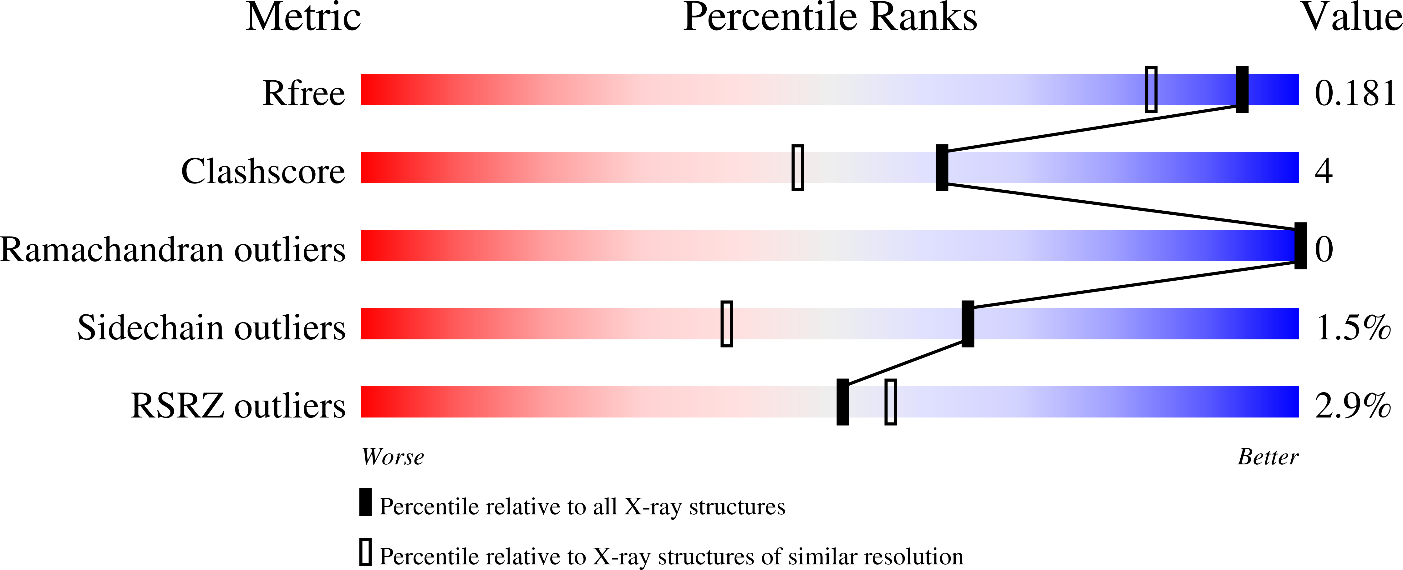
  • R-Value Free: 0.180 
  • R-Value Work: 0.162 
  • R-Value Observed: 0.163 

Starting Model: experimental
View more details

wwPDB Validation   3D Report Full Report


This is version 1.3 of the entry. See complete history


Literature

A Complete Structural Description of the Catalytic Cycle of Yeast Pyrophosphatase.

Oksanen, E.Ahonen, A.K.Tuominen, H.Tuominen, V.Lahti, R.Goldman, A.Heikinheimo, P.

(2007) Biochemistry 46: 1228-1239

  • DOI: https://doi.org/10.1021/bi0619977
  • Primary Citation of Related Structures:  
    2IHP, 2IK0, 2IK1, 2IK2, 2IK4, 2IK6, 2IK7, 2IK9

  • PubMed Abstract: 

    We have determined the structures of the wild type and seven active site variants of yeast inorganic pyrophosphatase (PPase) in the presence of Mg2+ and phosphate, providing the first complete structural description of its catalytic cycle. PPases catalyze the hydrolysis of pyrophosphate and require four divalent metal cations for catalysis; magnesium provides the highest activity. The crystal form chosen contains two monomers in the asymmetric unit, corresponding to distinct catalytic intermediates. In the "closed" wild-type active site, one of the two product phosphates has already dissociated, while the D115E variant "open" conformation is of the hitherto unobserved two-phosphate and two-"bridging" water active site. The mutations affect metal binding and the hydrogen bonding network in the active site, allowing us to explain the effects of mutations. For instance, in Y93F, F93 binds in a cryptic hydrophobic pocket in the absence of substrate, preserving hydrogen bonding in the active site and leading to relatively small changes in solution properties. This is not true in the presence of substrate, when F93 is forced back into the active site. Such subtle changes underline how low the energy barriers are between thermodynamically favorable conformations of the enzyme. The structures also allow us to associate metal binding constants to specific sites. Finally, the wild type and the D152E variant contain a phosphate ion adjacent to the active site, showing for the first time how product is released through a channel of flexible cationic side chains.


  • Organizational Affiliation

    Structural Biology and Biophysics, Institute of Biotechnology, P.O. Box 65, University of Helsinki, FIN-00014 Helsinki, Finland.


Macromolecules
Find similar proteins by:  (by identity cutoff)  |  3D Structure
Entity ID: 1
MoleculeChains Sequence LengthOrganismDetailsImage
Inorganic pyrophosphatase
A, B
286Saccharomyces cerevisiaeMutation(s): 0 
Gene Names: IPP1
EC: 3.6.1.1
UniProt
Find proteins for P00817 (Saccharomyces cerevisiae (strain ATCC 204508 / S288c))
Explore P00817 
Go to UniProtKB:  P00817
Entity Groups  
Sequence Clusters30% Identity50% Identity70% Identity90% Identity95% Identity100% Identity
UniProt GroupP00817
Sequence Annotations
Expand
  • Reference Sequence
Small Molecules
Ligands 3 Unique
IDChains Name / Formula / InChI Key2D Diagram3D Interactions
MES
Query on MES

Download Ideal Coordinates CCD File 
F [auth A]2-(N-MORPHOLINO)-ETHANESULFONIC ACID
C6 H13 N O4 S
SXGZJKUKBWWHRA-UHFFFAOYSA-N
PO4
Query on PO4

Download Ideal Coordinates CCD File 
E [auth A],
J [auth B]
PHOSPHATE ION
O4 P
NBIIXXVUZAFLBC-UHFFFAOYSA-K
MG
Query on MG

Download Ideal Coordinates CCD File 
C [auth A],
D [auth A],
G [auth B],
H [auth B],
I [auth B]
MAGNESIUM ION
Mg
JLVVSXFLKOJNIY-UHFFFAOYSA-N
Experimental Data & Validation

Experimental Data

  • Method: X-RAY DIFFRACTION
  • Resolution: 1.50 Å
  • R-Value Free: 0.180 
  • R-Value Work: 0.162 
  • R-Value Observed: 0.163 
  • Space Group: P 1 21 1
Unit Cell:
Length ( Å )Angle ( ˚ )
a = 51.915α = 90
b = 93.549β = 99.66
c = 69.168γ = 90
Software Package:
Software NamePurpose
REFMACrefinement
PDB_EXTRACTdata extraction
DENZOdata reduction
CCP4data scaling
MOLREPphasing

Structure Validation

View Full Validation Report


Entry History 

Deposition Data

Revision History  (Full details and data files)

  • Version 1.0: 2007-02-13
    Type: Initial release
  • Version 1.1: 2008-05-01
    Changes: Version format compliance
  • Version 1.2: 2011-07-13
    Changes: Version format compliance
  • Version 1.3: 2023-08-30
    Changes: Data collection, Database references, Derived calculations, Refinement description