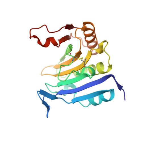
The structure of eukaryotic translation initiation factor-4E from wheat reveals a novel disulfide bond.
Monzingo, A.F., Dhaliwal, S., Dutt-Chaudhuri, A., Lyon, A., Sadow, J.H., Hoffman, D.W., Robertus, J.D., Browning, K.S.(2007) Plant Physiol 143: 1504-1518
- PubMed: 17322339 Search on PubMedSearch on PubMed Central
- DOI: https://doi.org/10.1104/pp.106.093146
- Primary Citation Related Structures:
2IDR, 2IDV - PubMed Abstract:
Eukaryotic translation initiation factor-4E (eIF4E) recognizes and binds the m(7) guanosine nucleotide at the 5' end of eukaryotic messenger RNAs; this protein-RNA interaction is an essential step in the initiation of protein synthesis. The structure of eIF4E from wheat (Triticum aestivum) was investigated using a combination of x-ray crystallography and nuclear magnetic resonance (NMR) methods. The overall fold of the crystallized protein was similar to eIF4E from other species, with eight beta-strands, three alpha-helices, and three extended loops. Surprisingly, the wild-type protein did not crystallize with m(7)GTP in its binding site, despite the ligand being present in solution; conformational changes in the cap-binding loops created a large cavity at the usual cap-binding site. The eIF4E crystallized in a dimeric form with one of the cap-binding loops of one monomer inserted into the cavity of the other. The protein also contained an intramolecular disulfide bridge between two cysteines (Cys) that are conserved only in plants. A Cys-to-serine mutant of wheat eIF4E, which lacked the ability to form the disulfide, crystallized with m(7)GDP in its binding pocket, with a structure similar to that of the eIF4E-cap complex of other species. NMR spectroscopy was used to show that the Cys that form the disulfide in the crystal are reduced in solution but can be induced to form the disulfide under oxidizing conditions. The observation that the disulfide-forming Cys are conserved in plants raises the possibility that their oxidation state may have a role in regulating protein function. NMR provided evidence that in oxidized eIF4E, the loop that is open in the ligand-free crystal dimer is relatively flexible in solution. An NMR-based binding assay showed that the reduced wheat eIF4E, the oxidized form with the disulfide, and the Cys-to-serine mutant protein each bind m(7)GTP in a similar and labile manner, with dissociation rates in the range of 20 to 100 s(-1).
- Department of Chemistry, University of Texas, Austin, Texas 78712, USA.
Organizational Affiliation: