2ID8 | pdb_00002id8

Crystal structure of Proteinase K


Experimental Data Snapshot

  • Method: X-RAY DIFFRACTION
  • Resolution: 1.27 Å
  • R-Value Free: 
    0.159 (Depositor) 
  • R-Value Work: 
    0.133 (DCC) 
  • R-Value Observed: 
    0.124 (Depositor) 

wwPDB Validation 3D Report Full Report

Validation slider image for 2ID8

Ligand Structure Quality Assessment 


This is version 1.3 of the entry. See complete history

Literature

What can be done with a good crystal and an accurate beamline?

Wang, J.Dauter, M.Dauter, Z.

(2006) Acta Crystallogr D Biol Crystallogr 62: 1475-1483

  • DOI: https://doi.org/10.1107/S0907444906038534
  • Primary Citation Related Structures: 
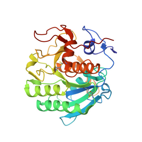
    2ID8

  • PubMed Abstract: 

    X-ray single-wavelength anomalous diffraction (SAD) data from a crystal of proteinase K were collected using synchrotron radiation of 0.98 A wavelength at SER-CAT 22-ID beamline, Advanced Photon Source, Argonne National Laboratory. At this wavelength, the expected Bijvoet ratio resulting from the presence of one calcium, one chloride and ten S atoms in the 279-residue protein is extremely small at approximately 0.46%. The direct-methods program SHELXD located 11 anomalous sites using data truncated to 2 A resolution. SHELXE was used to produce an easily interpretable electron-density map. This study shows that an accurate beamline and a good-quality crystal provide the possibility of successfully using a very weak anomalous signal of sulfur measured at a short wavelength for phasing a protein structure, even if a small degree of radiation damage is present.


  • Organizational Affiliation
    • SAIC-Frederick Inc., Basic Research Program, Argonne National Laboratory, Argonne, IL 60439, USA.

Macromolecule Content 

  • Total Structure Weight: 29.37 kDa 
  • Atom Count: 2,332 
  • Modeled Residue Count: 279 
  • Deposited Residue Count: 279 
  • Unique protein chains: 1

Macromolecules

Find similar proteins by:|  3D Structure
Entity ID: 1
MoleculeChains  Sequence LengthOrganismDetailsImage
Proteinase K279Parengyodontium albumMutation(s): 1 
EC: 3.4.21.64
UniProt
Find proteins for P06873 (Parengyodontium album)
Explore P06873 
Go to UniProtKB:  P06873
Entity Groups
Sequence Clusters30% Identity50% Identity70% Identity90% Identity95% Identity100% Identity
UniProt GroupP06873
Sequence Annotations
Expand
Reference Sequence

Small Molecules

Ligands 4 Unique
IDChains Name / Formula / InChI Key2D Diagram3D Interactions
2DB

Query on 2DB


Download:Ideal Coordinates CCD File
G [auth A](S)-(2,3-DIHYDROXYPROPOXY)TRIHYDROXYBORATE
C3 H10 B O6
KLKDDPLTQSQDLA-VKHMYHEASA-N
NO3

Query on NO3


Download:Ideal Coordinates CCD File
C [auth A],
D [auth A],
E [auth A]
NITRATE ION
N O3
NHNBFGGVMKEFGY-UHFFFAOYSA-N
CA

Query on CA


Download:Ideal Coordinates CCD File
B [auth A]CALCIUM ION
Ca
BHPQYMZQTOCNFJ-UHFFFAOYSA-N
CL

Query on CL


Download:Ideal Coordinates CCD File
F [auth A]CHLORIDE ION
Cl
VEXZGXHMUGYJMC-UHFFFAOYSA-M

Experimental Data & Validation

Experimental Data

  • Method: X-RAY DIFFRACTION
  • Resolution: 1.27 Å
  • R-Value Free:  0.159 (Depositor) 
  • R-Value Work:  0.133 (DCC) 
  • R-Value Observed: 0.124 (Depositor) 
Space Group: P 43 21 2
Unit Cell:
Length ( Å )Angle ( ˚ )
a = 67.55α = 90
b = 67.55β = 90
c = 106.88γ = 90
Software Package:
Software NamePurpose
SHELXmodel building
SHELXL-97refinement
SERGUIdata collection
HKL-2000data reduction
HKL-2000data scaling
SHELXDphasing
SHELXEmodel building

Structure Validation

View Full Validation Report


Ligand Structure Quality Assessment 


Entry History 

Deposition Data

Revision History  (Full details and data files)

  • Version 1.0: 2006-10-10
    Type: Initial release
  • Version 1.1: 2008-05-01
    Changes: Version format compliance
  • Version 1.2: 2011-07-13
    Changes: Version format compliance
  • Version 1.3: 2024-10-30
    Changes: Data collection, Database references, Derived calculations, Structure summary