2I7C | pdb_00002i7c

The crystal structure of spermidine synthase from p. falciparum in complex with AdoDATO


Experimental Data Snapshot

  • Method: X-RAY DIFFRACTION
  • Resolution: 1.71 Å
  • R-Value Free: 
    0.194 (Depositor), 0.192 (DCC) 
  • R-Value Work: 
    0.175 (Depositor), 0.173 (DCC) 
  • R-Value Observed: 
    0.176 (Depositor) 

Starting Model: experimental
View more details

wwPDB Validation 3D Report Full Report

Validation slider image for 2I7C

Ligand Structure Quality Assessment 


This is version 1.3 of the entry. See complete history

Literature

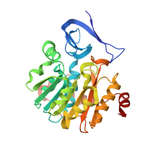
Crystal structure of Plasmodium falciparum spermidine synthase in complex with the substrate decarboxylated S-adenosylmethionine and the potent inhibitors 4MCHA and AdoDATO.

Dufe, V.T.Qiu, W.Muller, I.B.Hui, R.Walter, R.D.Al-Karadaghi, S.

(2007) J Mol Biology 373: 167-177

  • DOI: https://doi.org/10.1016/j.jmb.2007.07.053
  • Primary Citation Related Structures: 
    2I7C, 2PSS, 2PT6, 2PT9

  • PubMed Abstract: 

    Plasmodium falciparum is the causative agent of the most severe type of malaria, a life-threatening disease affecting the lives of over three billion people. Factors like widespread resistance against available drugs and absence of an effective vaccine are seriously compounding control of the malaria parasite. Thus, there is an urgent need for the identification and validation of new drug targets. The enzymes of the polyamine biosynthesis pathway have been suggested as possible targets for the treatment of malaria. One of these enzymes is spermidine synthase (SPDS, putrescine aminopropyltransferase), which catalyzes the transfer of an aminopropyl moiety from decarboxylated S-adenosylmethionine (dcAdoMet) to putrescine, leading to the formation of spermidine and 5'-methylthioadenosine. Here we present the three-dimensional structure of P. falciparum spermidine synthase (pfSPDS) in apo form, in complex with dcAdoMet and two inhibitors, S-adenosyl-1,8-diamino-3-thio-octane (AdoDATO) and trans-4-methylcyclohexylamine (4MCHA). The results show that binding of dcAdoMet to pfSPDS stabilizes the conformation of the flexible gatekeeper loop of the enzyme and affects the conformation of the active-site amino acid residues, preparing the protein for binding of the second substrate. The complexes of AdoDATO and 4MCHA with pfSPDS reveal the mode of interactions of these compounds with the enzyme. While AdoDATO essentially fills the entire active-site pocket, 4MCHA only occupies part of it, which suggests that simple modifications of this compound may yield more potent inhibitors of pfSPDS.


  • Organizational Affiliation
    • Department of Molecular Biophysics, Center for Molecular Protein Science, Lund University, S-221 00 Lund, Sweden.

Macromolecule Content 

  • Total Structure Weight: 98.71 kDa 
  • Atom Count: 7,559 
  • Modeled Residue Count: 842 
  • Deposited Residue Count: 849 
  • Unique protein chains: 1

Macromolecules

Find similar proteins by:|  3D Structure
Entity ID: 1
MoleculeChains  Sequence LengthOrganismDetailsImage
Spermidine synthase
A, B, C
283Plasmodium falciparum 3D7Mutation(s): 0 
EC: 2.5.1.16
UniProt
Find proteins for Q8II73 (Plasmodium falciparum (isolate 3D7))
Explore Q8II73 
Go to UniProtKB:  Q8II73
Entity Groups
Sequence Clusters30% Identity50% Identity70% Identity90% Identity95% Identity100% Identity
UniProt GroupQ8II73
Sequence Annotations
Expand
Reference Sequence

Small Molecules

Ligands 3 Unique
IDChains Name / Formula / InChI Key2D Diagram3D Interactions
AAT

Query on AAT


Download:Ideal Coordinates CCD File
D [auth A],
G [auth B],
J [auth C]
S-ADENOSYL-1,8-DIAMINO-3-THIOOCTANE
C18 H29 N7 O3 S
SUUGLGYBZXSJAA-SCFUHWHPSA-N
1PG

Query on 1PG


Download:Ideal Coordinates CCD File
E [auth A],
H [auth B]
2-(2-{2-[2-(2-METHOXY-ETHOXY)-ETHOXY]-ETHOXY}-ETHOXY)-ETHANOL
C11 H24 O6
SLNYBUIEAMRFSZ-UHFFFAOYSA-N
GOL

Query on GOL


Download:Ideal Coordinates CCD File
F [auth A],
I [auth B],
K [auth C]
GLYCEROL
C3 H8 O3
PEDCQBHIVMGVHV-UHFFFAOYSA-N

Experimental Data & Validation

Experimental Data

  • Method: X-RAY DIFFRACTION
  • Resolution: 1.71 Å
  • R-Value Free:  0.194 (Depositor), 0.192 (DCC) 
  • R-Value Work:  0.175 (Depositor), 0.173 (DCC) 
  • R-Value Observed: 0.176 (Depositor) 
Space Group: C 1 2 1
Unit Cell:
Length ( Å )Angle ( ˚ )
a = 198.168α = 90
b = 134.685β = 95.44
c = 48.5γ = 90
Software Package:
Software NamePurpose
REFMACrefinement
HKL-2000data collection
HKL-2000data reduction
HKL-2000data scaling

Structure Validation

View Full Validation Report


Ligand Structure Quality Assessment 


Entry History 

Revision History  (Full details and data files)

  • Version 1.0: 2006-09-12
    Type: Initial release
  • Version 1.1: 2008-05-01
    Changes: Version format compliance
  • Version 1.2: 2011-07-13
    Changes: Advisory, Source and taxonomy, Version format compliance
  • Version 1.3: 2023-08-30
    Changes: Data collection, Database references, Derived calculations, Refinement description