2I4A | pdb_00002i4a

Crystal structure of thioredoxin from the acidophile Acetobacter aceti


Experimental Data Snapshot

  • Method: X-RAY DIFFRACTION
  • Resolution: 1.00 Å
  • R-Value Free: 
    0.158 (Depositor), 0.164 (DCC) 
  • R-Value Work: 
    0.133 (Depositor), 0.143 (DCC) 
  • R-Value Observed: 
    0.135 (Depositor) 

Starting Model: experimental
View more details

wwPDB Validation 3D Report Full Report

Validation slider image for 2I4A

This is version 1.4 of the entry. See complete history

Literature

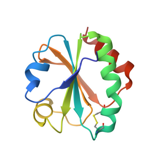
Atomic-resolution crystal structure of thioredoxin from the acidophilic bacterium Acetobacter aceti.

Starks, C.M.Francois, J.A.Macarthur, K.M.Heard, B.Z.Kappock, T.J.

(2007) Protein Sci 16: 92-98

  • DOI: https://doi.org/10.1110/ps.062519707
  • Primary Citation Related Structures: 
    2I4A

  • PubMed Abstract: 

    The crystal structure of thioredoxin (AaTrx) from the acetic acid bacterium Acetobacter aceti was determined at 1 A resolution. This is currently the highest resolution crystal structure available for any thioredoxin. Thioredoxins facilitate thiol-disulfide exchange, a process that is expected to be slow at the low pH values encountered in the A. aceti cytoplasm. Despite the apparent need to function at low pH, neither the active site nor the surface charge distribution of AaTrx is notably different from that of Escherichia coli thioredoxin. Apparently the ancestral thioredoxin was sufficiently stable for use in A. aceti or the need to interact with multiple targets constrained the variation of surface residues. The AaTrx structure presented here provides a clear view of all ionizable protein moieties and waters, a first step in understanding how thiol-disulfide exchange might occur in a low pH cytoplasm, and is a basis for biophysical studies of the mechanism of acid-mediated unfolding. The high resolution of this structure should be useful for computational studies of thioredoxin function, protein structure and dynamics, and side-chain ionization.


  • Organizational Affiliation
    • Department of Chemistry, Washington University in Saint Louis, Missouri 63130, USA.

Macromolecule Content 

  • Total Structure Weight: 11.65 kDa 
  • Atom Count: 1,004 
  • Modeled Residue Count: 107 
  • Deposited Residue Count: 107 
  • Unique protein chains: 1

Macromolecules

Find similar proteins by:|  3D Structure
Entity ID: 1
MoleculeChains  Sequence LengthOrganismDetailsImage
thioredoxin107Acetobacter acetiMutation(s): 0 

Small Molecules

Ligands 1 Unique
IDChains Name / Formula / InChI Key2D Diagram3D Interactions
BME

Query on BME


Download:Ideal Coordinates CCD File
B [auth A]BETA-MERCAPTOETHANOL
C2 H6 O S
DGVVWUTYPXICAM-UHFFFAOYSA-N

Experimental Data & Validation

Experimental Data

  • Method: X-RAY DIFFRACTION
  • Resolution: 1.00 Å
  • R-Value Free:  0.158 (Depositor), 0.164 (DCC) 
  • R-Value Work:  0.133 (Depositor), 0.143 (DCC) 
  • R-Value Observed: 0.135 (Depositor) 
Space Group: P 21 21 21
Unit Cell:
Length ( Å )Angle ( ˚ )
a = 33.94α = 90
b = 42.24β = 90
c = 63.67γ = 90
Software Package:
Software NamePurpose
REFMACrefinement
JBluIce-EPICSdata collection
d*TREKdata reduction
d*TREKdata scaling
MOLREPphasing

Structure Validation

View Full Validation Report


Entry History 

Deposition Data

Revision History  (Full details and data files)

  • Version 1.0: 2007-01-09
    Type: Initial release
  • Version 1.1: 2008-05-01
    Changes: Version format compliance
  • Version 1.2: 2011-07-13
    Changes: Version format compliance
  • Version 1.3: 2017-10-18
    Changes: Refinement description
  • Version 1.4: 2023-08-30
    Changes: Data collection, Database references, Derived calculations, Refinement description