2HXW | pdb_00002hxw

Crystal Structure of Peb3 from Campylobacter jejuni


Experimental Data Snapshot

  • Method: X-RAY DIFFRACTION
  • Resolution: 1.60 Å
  • R-Value Free: 
    0.212 (Depositor), 0.217 (DCC) 
  • R-Value Work: 
    0.187 (Depositor) 
  • R-Value Observed: 
    0.188 (Depositor) 

wwPDB Validation 3D Report Full Report

Validation slider image for 2HXW

This is version 1.4 of the entry. See complete history

Literature

Structural context for protein N-glycosylation in bacteria: The structure of PEB3, an adhesin from Campylobacter jejuni.

Rangarajan, E.S.Bhatia, S.Watson, D.C.Munger, C.Cygler, M.Matte, A.Young, N.M.

(2007) Protein Sci 16: 990-995

  • DOI: https://doi.org/10.1110/ps.062737507
  • Primary Citation Related Structures: 
    2HXW

  • PubMed Abstract: 

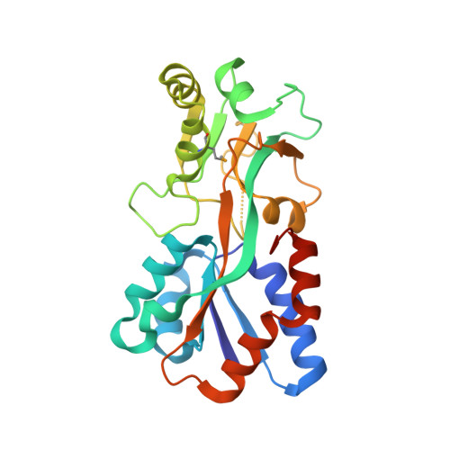
    Campylobacter jejuni is unusual among bacteria in possessing a eukaryotic-like system for N-linked protein glycosylation at Asn residues in sequons of the type Asp/Glu-Xaa-Asn-Xaa-Ser/Thr. However, little is known about the structural context of the glycosylated sequons, limiting the design of novel recombinant glycoproteins. To obtain more information on sequon structure, we have determined the crystal structure of the PEB3 (Cj0289c) dimer. PEB3 has the class II periplasmic-binding protein fold, with each monomer having two domains with a ligand-binding site containing citrate located between them, and overall resembles molybdate- and sulfate-binding proteins. The sequon around Asn90 is located within a surface-exposed loop joining two structural elements. The three key residues are well exposed on the surface; hence, they may be accessible to the PglB oligosaccharyltransferase in the folded state.


  • Organizational Affiliation
    • Department of Biochemistry, McGill University, Montreal, Quebec H3G 1Y6, Canada.

Macromolecule Content 

  • Total Structure Weight: 53.46 kDa 
  • Atom Count: 4,003 
  • Modeled Residue Count: 459 
  • Deposited Residue Count: 474 
  • Unique protein chains: 1

Macromolecules

Find similar proteins by:|  3D Structure
Entity ID: 1
MoleculeChains  Sequence LengthOrganismDetailsImage
Major antigenic peptide PEB3
A, B
237Campylobacter jejuniMutation(s): 1 
Gene Names: peb3
UniProt
Find proteins for Q0PBL7 (Campylobacter jejuni subsp. jejuni serotype O:2 (strain ATCC 700819 / NCTC 11168))
Explore Q0PBL7 
Go to UniProtKB:  Q0PBL7
Entity Groups
Sequence Clusters30% Identity50% Identity70% Identity90% Identity95% Identity100% Identity
UniProt GroupQ0PBL7
Sequence Annotations
Expand
Reference Sequence

Small Molecules

Modified Residues  1 Unique
IDChains TypeFormula2D DiagramParent
MSE
Query on MSE
A, B
L-PEPTIDE LINKINGC5 H11 N O2 SeMET

Experimental Data & Validation

Experimental Data

  • Method: X-RAY DIFFRACTION
  • Resolution: 1.60 Å
  • R-Value Free:  0.212 (Depositor), 0.217 (DCC) 
  • R-Value Work:  0.187 (Depositor) 
  • R-Value Observed: 0.188 (Depositor) 
Space Group: P 1 21 1
Unit Cell:
Length ( Å )Angle ( ˚ )
a = 46.472α = 90
b = 101.672β = 108.98
c = 59.099γ = 90
Software Package:
Software NamePurpose
REFMACrefinement
HKL-2000data collection
HKL-2000data reduction
HKL-2000data scaling
SHELXSphasing

Structure Validation

View Full Validation Report


Entry History 

Revision History  (Full details and data files)

  • Version 1.0: 2007-05-01
    Type: Initial release
  • Version 1.1: 2008-05-01
    Changes: Version format compliance
  • Version 1.2: 2011-07-13
    Changes: Version format compliance
  • Version 1.3: 2022-12-21
    Changes: Database references, Derived calculations
  • Version 1.4: 2024-10-16
    Changes: Data collection, Structure summary