2HX2 | pdb_00002hx2

Bovine eNOS heme domain complexed with (4S)-N-{4-Amino-5-[(2-aminoethyl)-hydroxyamino]-pentyl}-N'-nitroguanidine


Experimental Data Snapshot

  • Method: X-RAY DIFFRACTION
  • Resolution: 1.95 Å
  • R-Value Free: 
    0.213 (Depositor), 0.209 (DCC) 
  • R-Value Work: 
    0.180 (Depositor), 0.177 (DCC) 
  • R-Value Observed: 
    0.180 (Depositor) 

wwPDB Validation 3D Report Full Report

Validation slider image for 2HX2

Ligand Structure Quality Assessment 


This is version 1.3 of the entry. See complete history

Literature

Structure-Based Design and Synthesis of N(omega)-Nitro-l-Arginine-Containing Peptidomimetics as Selective Inhibitors of Neuronal Nitric Oxide Synthase. Displacement of the Heme Structural Water.

Seo, J.Igarashi, J.Li, H.Martasek, P.Roman, L.J.Poulos, T.L.Silverman, R.B.

(2007) J Med Chem 50: 2089-2099

  • DOI: https://doi.org/10.1021/jm061305c
  • Primary Citation Related Structures: 
    2HX2, 2HX3, 2HX4

  • PubMed Abstract: 

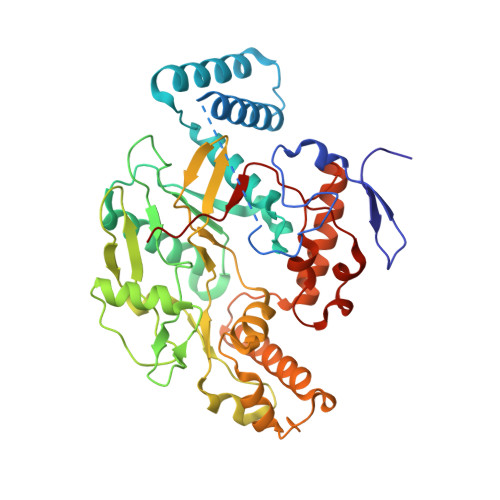
    The neuronal isoform of nitric oxide synthase (nNOS), the enzyme responsible for the production of nitric oxide in the central nervous system, represents an attractive target for the treatment of various neurodegenerative disorders. X-ray crystal structures of complexes of nNOS with two nNOS-selective inhibitors, (4S)-N-{4-amino-5-[(2-aminoethylamino]pentyl}-N'-nitroguanidine (1) and 4-N-(Nomega-nitro-l-argininyl)-trans-4-amino-l-proline amide (2), led to the discovery of a conserved structural water molecule that was hydrogen bonded between the two heme propionates and the inhibitors (Figure 2). On the basis of this observation, we hypothesized that by attaching a hydrogen bond donor group to the amide nitrogen of 2 or to the secondary amine nitrogen of 1, the inhibitor molecules could displace the structural water molecule and obtain a direct interaction with the heme cofactor. To test this hypothesis, peptidomimetic analogues 3-5, which have either an N-hydroxyl (3 and 5) or N-amino (4) donor group, were designed and synthesized. X-ray crystal structures of nNOS with inhibitors 3 and 5 bound verified that the N-hydroxyl group had, indeed, displaced the structural water molecule and provided a direct interaction with the heme propionate moiety (Figures 5 and 6). Surprisingly, in vitro activity assay results indicated that the addition of a hydroxyl group (3) only increased the potency slightly against the neuronal isoform over the parent compound (1). Rationalizations for the small increase in potency are consistent with other changes in the crystal structures.


  • Organizational Affiliation
    • Department of Chemistry, Northwestern University, Evanston, Illinois 60208-3113, USA.

Macromolecule Content 

  • Total Structure Weight: 96.71 kDa 
  • Atom Count: 7,132 
  • Modeled Residue Count: 805 
  • Deposited Residue Count: 832 
  • Unique protein chains: 1

Macromolecules

Find similar proteins by:|  3D Structure
Entity ID: 1
MoleculeChains  Sequence LengthOrganismDetailsImage
Nitric-oxide synthase
A, B
416Bos taurusMutation(s): 0 
Gene Names: NOS3
EC: 1.14.13.39
UniProt
Find proteins for P29473 (Bos taurus)
Explore P29473 
Go to UniProtKB:  P29473
Entity Groups
Sequence Clusters30% Identity50% Identity70% Identity90% Identity95% Identity100% Identity
UniProt GroupP29473
Sequence Annotations
Expand
Reference Sequence

Small Molecules

Ligands 7 Unique
IDChains Name / Formula / InChI Key2D Diagram3D Interactions
HEM

Query on HEM


Download:Ideal Coordinates CCD File
G [auth A],
N [auth B]
PROTOPORPHYRIN IX CONTAINING FE
C34 H32 Fe N4 O4
KABFMIBPWCXCRK-RGGAHWMASA-L
3HX

Query on 3HX


Download:Ideal Coordinates CCD File
I [auth A],
P [auth B]
(4S)-N-{4-AMINO-5-[(2-AMINOETHYL)(HYDROXYAMINO]-PENTYL}-N'-NITROGUANIDINE
C8 H21 N7 O3
HUOGRSGQHCPAGO-ZETCQYMHSA-N
H4B

Query on H4B


Download:Ideal Coordinates CCD File
H [auth A],
O [auth B]
5,6,7,8-TETRAHYDROBIOPTERIN
C9 H15 N5 O3
FNKQXYHWGSIFBK-RPDRRWSUSA-N
CAC

Query on CAC


Download:Ideal Coordinates CCD File
C [auth A],
K [auth B]
CACODYLATE ION
C2 H6 As O2
OGGXGZAMXPVRFZ-UHFFFAOYSA-M
GOL

Query on GOL


Download:Ideal Coordinates CCD File
J [auth A],
Q [auth B]
GLYCEROL
C3 H8 O3
PEDCQBHIVMGVHV-UHFFFAOYSA-N
ZN

Query on ZN


Download:Ideal Coordinates CCD File
F [auth A]ZINC ION
Zn
PTFCDOFLOPIGGS-UHFFFAOYSA-N
ACT

Query on ACT


Download:Ideal Coordinates CCD File
D [auth A],
E [auth A],
L [auth B],
M [auth B]
ACETATE ION
C2 H3 O2
QTBSBXVTEAMEQO-UHFFFAOYSA-M

Experimental Data & Validation

Experimental Data

  • Method: X-RAY DIFFRACTION
  • Resolution: 1.95 Å
  • R-Value Free:  0.213 (Depositor), 0.209 (DCC) 
  • R-Value Work:  0.180 (Depositor), 0.177 (DCC) 
  • R-Value Observed: 0.180 (Depositor) 
Space Group: P 21 21 21
Unit Cell:
Length ( Å )Angle ( ˚ )
a = 57.82α = 90
b = 106.85β = 90
c = 156.35γ = 90
Software Package:
Software NamePurpose
CNSrefinement
HKL-2000data collection
HKL-2000data reduction
HKL-2000data scaling
CNSphasing

Structure Validation

View Full Validation Report


Ligand Structure Quality Assessment 


Entry History 

Deposition Data

Revision History  (Full details and data files)

  • Version 1.0: 2007-04-24
    Type: Initial release
  • Version 1.1: 2008-04-01
    Changes: Version format compliance
  • Version 1.2: 2011-07-13
    Changes: Derived calculations, Version format compliance
  • Version 1.3: 2024-02-14
    Changes: Data collection, Database references, Derived calculations