2HWW

Structure of PIN domain of human SMG6


Experimental Data Snapshot

  • Method: X-RAY DIFFRACTION
  • Resolution: 1.80 Å
  • R-Value Free: 0.267 
  • R-Value Work: 0.222 
  • R-Value Observed: 0.224 

Starting Model: experimental
View more details

wwPDB Validation   3D Report Full Report


This is version 1.3 of the entry. See complete history


Literature

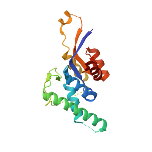
Structures of the PIN domains of SMG6 and SMG5 reveal a nuclease within the mRNA surveillance complex.

Glavan, F.Behm-Ansmant, I.Izaurralde, E.Conti, E.

(2006) EMBO J 25: 5117-5125

  • DOI: https://doi.org/10.1038/sj.emboj.7601377
  • Primary Citation of Related Structures:  
    2HWW, 2HWX, 2HWY

  • PubMed Abstract: 

    SMG6 and SMG5 are essential factors in nonsense-mediated mRNA decay, a conserved pathway that degrades mRNAs with premature translation termination codons. Both SMG5 and SMG6 have been predicted to contain a C-terminal PIN (PilT N-terminus) domain, present in proteins with ribonuclease activity. We have determined the structures of human SMG5 and SMG6 PIN domains. Although they share a similar overall fold related to ribonucleases of the RNase H family, they have local differences at the putative active site. SMG6 has the canonical triad of acidic residues that are crucial in RNase H for nuclease activity, while SMG5 lacks key catalytic residues. The structural differences are reflected at the functional level. Only the PIN domain of SMG6 has degradation activity on single-stranded RNA in vitro. This difference in catalytic activity is conserved in Drosophila, where an SMG6 with an inactive PIN domain inhibits NMD in a dominant-negative manner. Our findings suggest that the NMD machinery has intrinsic nuclease activity that is likely to contribute to the rapid decay of mRNAs that terminate translation prematurely.


  • Organizational Affiliation

    European Molecular Biology Laboratory, Heidelberg, Germany.


Macromolecules
Find similar proteins by:  (by identity cutoff)  |  3D Structure
Entity ID: 1
MoleculeChains Sequence LengthOrganismDetailsImage
Telomerase-binding protein EST1A
A, B, C
181Homo sapiensMutation(s): 0 
Gene Names: EST1A
UniProt & NIH Common Fund Data Resources
Find proteins for Q86US8 (Homo sapiens)
Explore Q86US8 
Go to UniProtKB:  Q86US8
PHAROS:  Q86US8
GTEx:  ENSG00000070366 
Entity Groups  
Sequence Clusters30% Identity50% Identity70% Identity90% Identity95% Identity100% Identity
UniProt GroupQ86US8
Sequence Annotations
Expand
  • Reference Sequence
Experimental Data & Validation

Experimental Data

  • Method: X-RAY DIFFRACTION
  • Resolution: 1.80 Å
  • R-Value Free: 0.267 
  • R-Value Work: 0.222 
  • R-Value Observed: 0.224 
  • Space Group: P 21 21 21
Unit Cell:
Length ( Å )Angle ( ˚ )
a = 36.879α = 90
b = 71.216β = 90
c = 181.273γ = 90
Software Package:
Software NamePurpose
REFMACrefinement
MOSFLMdata reduction
CCP4data scaling
PHASERphasing

Structure Validation

View Full Validation Report


Entry History 

Deposition Data

Revision History  (Full details and data files)

  • Version 1.0: 2006-11-14
    Type: Initial release
  • Version 1.1: 2008-05-01
    Changes: Version format compliance
  • Version 1.2: 2011-07-13
    Changes: Version format compliance
  • Version 1.3: 2023-08-30
    Changes: Data collection, Database references, Refinement description