2HW1

Crystal structure of human ketohexokinase complexed to different sugar molecules


Experimental Data Snapshot

  • Method: X-RAY DIFFRACTION
  • Resolution: 2.10 Å
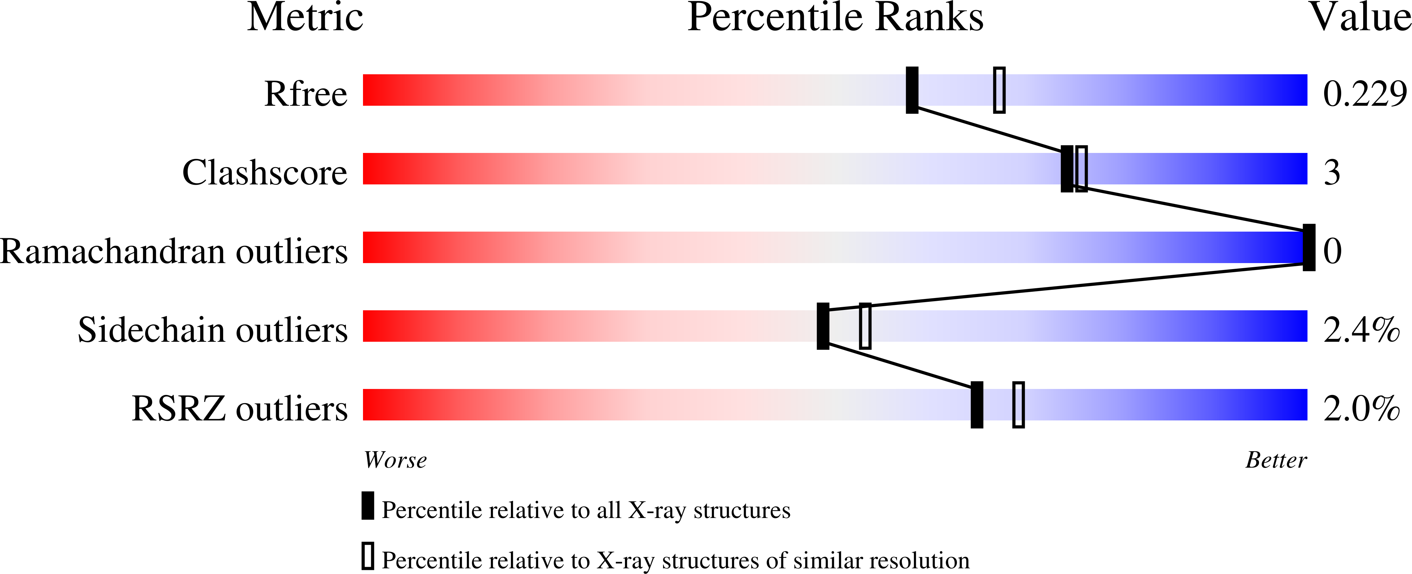
  • R-Value Free: 0.228 
  • R-Value Work: 0.193 
  • R-Value Observed: 0.194 

Starting Model: experimental
View more details

wwPDB Validation   3D Report Full Report


Ligand Structure Quality Assessment 


This is version 1.4 of the entry. See complete history


Literature

Structures of alternatively spliced isoforms of human ketohexokinase.

Trinh, C.H.Asipu, A.Bonthron, D.T.Phillips, S.E.

(2009) Acta Crystallogr D Biol Crystallogr 65: 201-211

  • DOI: https://doi.org/10.1107/S0907444908041115
  • Primary Citation of Related Structures:  
    2HQQ, 2HW1

  • PubMed Abstract: 

    A molecular understanding of the unique aspects of dietary fructose metabolism may be the key to understanding and controlling the current epidemic of fructose-related obesity, diabetes and related adverse metabolic states in Western populations. Fructose catabolism is initiated by its phosphorylation to fructose 1-phosphate, which is performed by ketohexokinase (KHK). Here, the crystal structures of the two alternatively spliced isoforms of human ketohexokinase, hepatic KHK-C and the peripheral isoform KHK-A, and of the ternary complex of KHK-A with the substrate fructose and AMP-PNP are reported. The structure of the KHK-A ternary complex revealed an active site with both the substrate fructose and the ATP analogue in positions ready for phosphorylation following a reaction mechanism similar to that of the pfkB family of carbohydrate kinases. Hepatic KHK deficiency causes the benign disorder essential fructosuria. The effects of the disease-causing mutations (Gly40Arg and Ala43Thr) have been modelled in the context of the KHK structure.


  • Organizational Affiliation

    Astbury Centre for Structural Molecular Biology, Institute of Molecular and Cellular Biology, University of Leeds, Leeds, England.


Macromolecules
Find similar proteins by:  (by identity cutoff)  |  3D Structure
Entity ID: 1
MoleculeChains Sequence LengthOrganismDetailsImage
Ketohexokinase298Homo sapiensMutation(s): 0 
Gene Names: KHK
EC: 2.7.1.3
UniProt & NIH Common Fund Data Resources
Find proteins for P50053 (Homo sapiens)
Explore P50053 
Go to UniProtKB:  P50053
PHAROS:  P50053
GTEx:  ENSG00000138030 
Entity Groups  
Sequence Clusters30% Identity50% Identity70% Identity90% Identity95% Identity100% Identity
UniProt GroupP50053
Sequence Annotations
Expand
  • Reference Sequence
Experimental Data & Validation

Experimental Data

  • Method: X-RAY DIFFRACTION
  • Resolution: 2.10 Å
  • R-Value Free: 0.228 
  • R-Value Work: 0.193 
  • R-Value Observed: 0.194 
  • Space Group: I 2 2 2
Unit Cell:
Length ( Å )Angle ( ˚ )
a = 61.264α = 90
b = 107.823β = 90
c = 146.679γ = 90
Software Package:
Software NamePurpose
REFMACrefinement
MOSFLMdata reduction
SCALAdata scaling
AMoREphasing

Structure Validation

View Full Validation Report


Ligand Structure Quality Assessment 


Entry History 

Deposition Data

Revision History  (Full details and data files)

  • Version 1.0: 2007-07-10
    Type: Initial release
  • Version 1.1: 2008-05-01
    Changes: Version format compliance
  • Version 1.2: 2011-07-13
    Changes: Non-polymer description, Version format compliance
  • Version 1.3: 2020-07-29
    Type: Remediation
    Reason: Carbohydrate remediation
    Changes: Data collection, Derived calculations, Structure summary
  • Version 1.4: 2023-08-30
    Changes: Data collection, Database references, Refinement description, Structure summary