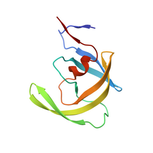
2HS2

Crystal structure of M46L mutant of HIV-1 protease complexed with TMC114 (darunavir)


Experimental Data Snapshot

  • Method: X-RAY DIFFRACTION
  • Resolution: 1.22 Å
  • R-Value Free: 0.195 
  • R-Value Work: 0.140 
  • R-Value Observed: 0.140 

wwPDB Validation   3D Report Full Report


Ligand Structure Quality Assessment 


This is version 1.4 of the entry. See complete history


Literature

Ultra-high Resolution Crystal Structure of HIV-1 Protease Mutant Reveals Two Binding Sites for Clinical Inhibitor TMC114.

Kovalevsky, A.Y.Liu, F.Leshchenko, S.Ghosh, A.K.Louis, J.M.Harrison, R.W.Weber, I.T.

(2006) J Mol Biol 363: 161-173

  • DOI: https://doi.org/10.1016/j.jmb.2006.08.007
  • Primary Citation of Related Structures:  
    2HS1, 2HS2

  • PubMed Abstract: 

    TMC114 (darunavir) is a promising clinical inhibitor of HIV-1 protease (PR) for treatment of drug resistant HIV/AIDS. We report the ultra-high 0.84 A resolution crystal structure of the TMC114 complex with PR containing the drug-resistant mutation V32I (PR(V32I)), and the 1.22 A resolution structure of a complex with PR(M46L). These structures show TMC114 bound at two distinct sites, one in the active-site cavity and the second on the surface of one of the flexible flaps in the PR dimer. Remarkably, TMC114 binds at these two sites simultaneously in two diastereomers related by inversion of the sulfonamide nitrogen. Moreover, the flap site is shaped to accommodate the diastereomer with the S-enantiomeric nitrogen rather than the one with the R-enantiomeric nitrogen. The existence of the second binding site and two diastereomers suggest a mechanism for the high effectiveness of TMC114 on drug-resistant HIV and the potential design of new inhibitors.


  • Organizational Affiliation

    Department of Biology, Molecular Basis of Disease, GA State University, Atlanta, GA 30303, USA.


Macromolecules
Find similar proteins by:  (by identity cutoff)  |  3D Structure
Entity ID: 1
MoleculeChains Sequence LengthOrganismDetailsImage
protease
A, B
99Human immunodeficiency virus 1Mutation(s): 6 
Gene Names: GAG
EC: 3.4.23.16
UniProt
Find proteins for Q7SSI0 (Human immunodeficiency virus 1)
Explore Q7SSI0 
Go to UniProtKB:  Q7SSI0
Entity Groups  
Sequence Clusters30% Identity50% Identity70% Identity90% Identity95% Identity100% Identity
UniProt GroupQ7SSI0
Sequence Annotations
Expand
  • Reference Sequence
Small Molecules
Binding Affinity Annotations 
IDSourceBinding Affinity
017 BindingDB:  2HS2 Ki: min: 0.01, max: 6.6 (nM) from 8 assay(s)
EC50: 1.6 (nM) from 1 assay(s)
Experimental Data & Validation

Experimental Data

  • Method: X-RAY DIFFRACTION
  • Resolution: 1.22 Å
  • R-Value Free: 0.195 
  • R-Value Work: 0.140 
  • R-Value Observed: 0.140 
  • Space Group: P 21 21 21
Unit Cell:
Length ( Å )Angle ( ˚ )
a = 28.913α = 90
b = 66.562β = 90
c = 93.147γ = 90
Software Package:
Software NamePurpose
SHELXmodel building
SHELXL-97refinement
HKL-2000data reduction
HKL-2000data scaling
SHELXphasing

Structure Validation

View Full Validation Report


Ligand Structure Quality Assessment 


Entry History 

Deposition Data

Revision History  (Full details and data files)

  • Version 1.0: 2006-10-03
    Type: Initial release
  • Version 1.1: 2008-04-01
    Changes: Version format compliance
  • Version 1.2: 2011-07-13
    Changes: Non-polymer description, Version format compliance
  • Version 1.3: 2021-10-20
    Changes: Database references, Derived calculations, Structure summary
  • Version 1.4: 2024-02-14
    Changes: Data collection