2HPD | pdb_00002hpd

CRYSTAL STRUCTURE OF HEMOPROTEIN DOMAIN OF P450BM-3, A PROTOTYPE FOR MICROSOMAL P450'S


Experimental Data Snapshot

  • Method: X-RAY DIFFRACTION
  • Resolution: 2.00 Å
  • R-Value Work: 
    0.167 (Depositor) 
  • R-Value Observed: 
    0.167 (Depositor) 

wwPDB Validation 3D Report Full Report

Validation slider image for 2HPD

This is version 1.3 of the entry. See complete history

Literature

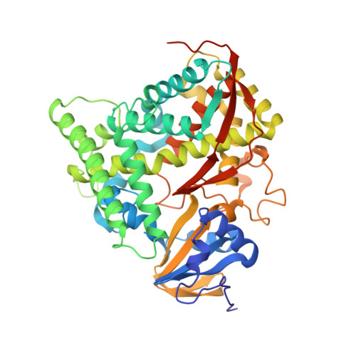
Crystal structure of hemoprotein domain of P450BM-3, a prototype for microsomal P450's.

Ravichandran, K.G.Boddupalli, S.S.Hasermann, C.A.Peterson, J.A.Deisenhofer, J.

(1993) Science 261: 731-736

  • DOI: https://doi.org/10.1126/science.8342039
  • Primary Citation Related Structures: 
    2HPD

  • PubMed Abstract: 

    Cytochrome P450BM-3, a bacterial fatty acid monoxygenase, resembles the eukaryotic microsomal P450's and their flavoprotein reductase in primary structure and function. The three-dimensional structure of the hemoprotein domain of P450BM-3 was determined by x-ray diffraction and refined to an R factor of 16.9 percent at 2.0 angstrom resolution. The structure consists of an alph and a beta domain. The active site heme is accessible through a long hydrophobic channel formed primarily by the beta domain and the B' and F helices of the alpha domain. The two molecules in the asymmetric unit differ in conformation around the substrate binding pocket. Substantial differences between P450BM-3 and P450cam, the only other P450 structure available, are observed around the substrate binding pocket and the regions important for redox partner binding. A general mechanism for proton transfer in P450's is also proposed.


  • Organizational Affiliation
    • Howard Hughes Medical Institute, University of Texas Southwestern Medical Center, Dallas 75235-9050.

Macromolecule Content 

  • Total Structure Weight: 108.83 kDa 
  • Atom Count: 7,897 
  • Modeled Residue Count: 914 
  • Deposited Residue Count: 942 
  • Unique protein chains: 1

Macromolecules

Find similar proteins by:|  3D Structure
Entity ID: 1
MoleculeChains  Sequence LengthOrganismDetailsImage
CYTOCHROME P450 BM-3
A, B
471Priestia megateriumMutation(s): 0 
EC: 1.14.14.1 (PDB Primary Data), 1.6.2.4 (UniProt)
UniProt
Find proteins for P14779 (Priestia megaterium (strain ATCC 14581 / DSM 32 / CCUG 1817 / JCM 2506 / NBRC 15308 / NCIMB 9376 / NCTC 10342 / NRRL B-14308 / VKM B-512 / Ford 19))
Explore P14779 
Go to UniProtKB:  P14779
Entity Groups
Sequence Clusters30% Identity50% Identity70% Identity90% Identity95% Identity100% Identity
UniProt GroupP14779
Sequence Annotations
Expand
Reference Sequence

Small Molecules

Ligands 1 Unique
IDChains Name / Formula / InChI Key2D Diagram3D Interactions
HEM

Query on HEM


Download:Ideal Coordinates CCD File
C [auth A],
D [auth B]
PROTOPORPHYRIN IX CONTAINING FE
C34 H32 Fe N4 O4
KABFMIBPWCXCRK-RGGAHWMASA-L

Experimental Data & Validation

Experimental Data

  • Method: X-RAY DIFFRACTION
  • Resolution: 2.00 Å
  • R-Value Work:  0.167 (Depositor) 
  • R-Value Observed: 0.167 (Depositor) 
Space Group: P 1 21 1
Unit Cell:
Length ( Å )Angle ( ˚ )
a = 59.4α = 90
b = 154β = 94.7
c = 62.2γ = 90
Software Package:
Software NamePurpose
X-PLORmodel building
X-PLORrefinement
X-PLORphasing

Structure Validation

View Full Validation Report


Entry History 

Deposition Data

Revision History  (Full details and data files)

  • Version 1.0: 1993-10-31
    Type: Initial release
  • Version 1.1: 2008-03-24
    Changes: Version format compliance
  • Version 1.2: 2011-07-13
    Changes: Version format compliance
  • Version 1.3: 2024-02-14
    Changes: Data collection, Database references, Derived calculations, Other