2HLD | pdb_00002hld

Crystal structure of yeast mitochondrial F1-ATPase


Experimental Data Snapshot

  • Method: X-RAY DIFFRACTION
  • Resolution: 2.80 Å
  • R-Value Free: 
    0.244 (Depositor), 0.337 (DCC) 
  • R-Value Work: 
    0.207 (Depositor) 
  • R-Value Observed: 
    0.207 (Depositor) 

Starting Model: experimental
View more details

wwPDB Validation 3D Report Full Report

Validation slider image for 2HLD

Ligand Structure Quality Assessment 


This is version 1.3 of the entry. See complete history

Literature

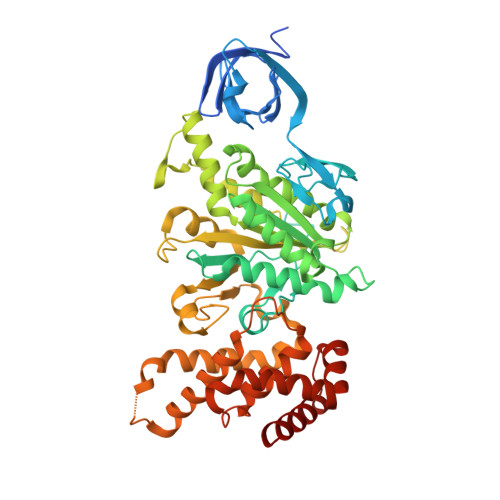
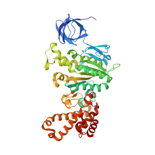
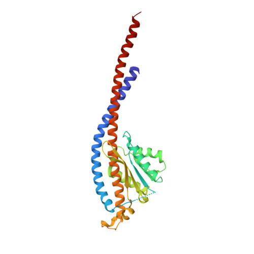
Novel features of the rotary catalytic mechanism revealed in the structure of yeast F(1) ATPase.

Kabaleeswaran, V.Puri, N.Walker, J.E.Leslie, A.G.Mueller, D.M.

(2006) EMBO J 25: 5433-5442

  • DOI: https://doi.org/10.1038/sj.emboj.7601410
  • Primary Citation Related Structures: 
    2HLD

  • PubMed Abstract: 

    The crystal structure of yeast mitochondrial F(1) ATPase contains three independent copies of the complex, two of which have similar conformations while the third differs in the position of the central stalk relative to the alpha(3)beta(3) sub-assembly. All three copies display very similar asymmetric features to those observed for the bovine enzyme, but the yeast F(1) ATPase structures provide novel information. In particular, the active site that binds ADP in bovine F(1) ATPase has an ATP analog bound and therefore this structure does not represent the ADP-inhibited form. In addition, one of the complexes binds phosphate in the nucleotide-free catalytic site, and comparison with other structures provides a picture of the movement of the phosphate group during initial binding and subsequent catalysis. The shifts in position of the central stalk between two of the three copies of yeast F(1) ATPase and when these structures are compared to those of the bovine enzyme give new insight into the conformational changes that take place during rotational catalysis.


  • Organizational Affiliation
    • Department of Biochemistry & Molecular Biology, The Chicago Medical School, Rosalind Franklin University of Medicine and Science, North Chicago, IL 60064, USA.

Macromolecule Content 

  • Total Structure Weight: 1,119.27 kDa 
  • Atom Count: 72,841 
  • Modeled Residue Count: 9,599 
  • Deposited Residue Count: 10,323 
  • Unique protein chains: 5

Macromolecules

Find similar proteins by:|  3D Structure
Entity ID: 1
MoleculeChains  Sequence LengthOrganismDetailsImage
ATP synthase alpha chain, mitochondrial
A, B, C, J, K
A, B, C, J, K, L, S, T, U
510Saccharomyces cerevisiaeMutation(s): 0 
EC: 3.6.3.14
Membrane Entity: Yes 
UniProt
Find proteins for P07251 (Saccharomyces cerevisiae (strain ATCC 204508 / S288c))
Explore P07251 
Go to UniProtKB:  P07251
Entity Groups
Sequence Clusters30% Identity50% Identity70% Identity90% Identity95% Identity100% Identity
UniProt GroupP07251
Sequence Annotations
Expand
Reference Sequence
Find similar proteins by:|  3D Structure
Entity ID: 2
MoleculeChains  Sequence LengthOrganismDetailsImage
ATP synthase beta chain, mitochondrial
D, E, F, M, N
D, E, F, M, N, O, V, W, X
478Saccharomyces cerevisiaeMutation(s): 0 
Gene Names: ATP2
EC: 3.6.3.14 (PDB Primary Data), 7.1.2.2 (UniProt)
Membrane Entity: Yes 
UniProt
Find proteins for P00830 (Saccharomyces cerevisiae (strain ATCC 204508 / S288c))
Explore P00830 
Go to UniProtKB:  P00830
Entity Groups
Sequence Clusters30% Identity50% Identity70% Identity90% Identity95% Identity100% Identity
UniProt GroupP00830
Sequence Annotations
Expand
Reference Sequence
Find similar proteins by:|  3D Structure
Entity ID: 3
MoleculeChains  Sequence LengthOrganismDetailsImage
ATP synthase gamma chain, mitochondrial
G, P, Y
278Saccharomyces cerevisiaeMutation(s): 0 
EC: 3.6.3.14
Membrane Entity: Yes 
UniProt
Find proteins for P38077 (Saccharomyces cerevisiae (strain ATCC 204508 / S288c))
Explore P38077 
Go to UniProtKB:  P38077
Entity Groups
Sequence Clusters30% Identity50% Identity70% Identity90% Identity95% Identity100% Identity
UniProt GroupP38077
Sequence Annotations
Expand
Reference Sequence
Find similar proteins by:|  3D Structure
Entity ID: 4
MoleculeChains  Sequence LengthOrganismDetailsImage
ATP synthase delta chain, mitochondrial
H, Q, Z
138Saccharomyces cerevisiaeMutation(s): 0 
EC: 3.6.3.14
Membrane Entity: Yes 
UniProt
Find proteins for Q12165 (Saccharomyces cerevisiae (strain ATCC 204508 / S288c))
Explore Q12165 
Go to UniProtKB:  Q12165
Entity Groups
Sequence Clusters30% Identity50% Identity70% Identity90% Identity95% Identity100% Identity
UniProt GroupQ12165
Sequence Annotations
Expand
Reference Sequence
Find similar proteins by:|  3D Structure
Entity ID: 5
MoleculeChains  Sequence LengthOrganismDetailsImage
ATP synthase epsilon chain, mitochondrialAA [auth 1],
I,
R
61Saccharomyces cerevisiaeMutation(s): 0 
EC: 3.6.3.14
Membrane Entity: Yes 
UniProt
Find proteins for P21306 (Saccharomyces cerevisiae (strain ATCC 204508 / S288c))
Explore P21306 
Go to UniProtKB:  P21306
Entity Groups
Sequence Clusters30% Identity50% Identity70% Identity90% Identity95% Identity100% Identity
UniProt GroupP21306
Sequence Annotations
Expand
Reference Sequence

Small Molecules

Ligands 3 Unique
IDChains Name / Formula / InChI Key2D Diagram3D Interactions
ANP

Query on ANP


Download:Ideal Coordinates CCD File
BB [auth U]
CA [auth A]
DB [auth V]
EA [auth B]
FB [auth X]
BB [auth U],
CA [auth A],
DB [auth V],
EA [auth B],
FB [auth X],
GA [auth C],
IA [auth D],
KA [auth F],
MA [auth J],
OA [auth K],
QA [auth L],
SA [auth M],
VA [auth O],
XA [auth S],
ZA [auth T]
PHOSPHOAMINOPHOSPHONIC ACID-ADENYLATE ESTER
C10 H17 N6 O12 P3
PVKSNHVPLWYQGJ-KQYNXXCUSA-N
PO4

Query on PO4


Download:Ideal Coordinates CCD File
TA [auth N]PHOSPHATE ION
O4 P
NBIIXXVUZAFLBC-UHFFFAOYSA-K
MG

Query on MG


Download:Ideal Coordinates CCD File
AB [auth U]
BA [auth A]
CB [auth V]
DA [auth B]
EB [auth X]
AB [auth U],
BA [auth A],
CB [auth V],
DA [auth B],
EB [auth X],
FA [auth C],
HA [auth D],
JA [auth F],
LA [auth J],
NA [auth K],
PA [auth L],
RA [auth M],
UA [auth O],
WA [auth S],
YA [auth T]
MAGNESIUM ION
Mg
JLVVSXFLKOJNIY-UHFFFAOYSA-N

Experimental Data & Validation

Experimental Data

  • Method: X-RAY DIFFRACTION
  • Resolution: 2.80 Å
  • R-Value Free:  0.244 (Depositor), 0.337 (DCC) 
  • R-Value Work:  0.207 (Depositor) 
  • R-Value Observed: 0.207 (Depositor) 
Space Group: P 1 21 1
Unit Cell:
Length ( Å )Angle ( ˚ )
a = 111.524α = 90
b = 294.132β = 101.67
c = 190.432γ = 90
Software Package:
Software NamePurpose
REFMACrefinement
MOSFLMdata reduction
CCP4data scaling
AMoREphasing

Structure Validation

View Full Validation Report


Ligand Structure Quality Assessment 


Entry History 

Deposition Data

Revision History  (Full details and data files)

  • Version 1.0: 2006-11-28
    Type: Initial release
  • Version 1.1: 2008-05-01
    Changes: Version format compliance
  • Version 1.2: 2011-07-13
    Changes: Advisory, Refinement description, Version format compliance
  • Version 1.3: 2023-08-30
    Changes: Data collection, Database references, Derived calculations, Refinement description