2HCJ | pdb_00002hcj

Trypsin-modified Elongation Factor Tu in complex with tetracycline


Experimental Data Snapshot

  • Method: X-RAY DIFFRACTION
  • Resolution: 2.12 Å
  • R-Value Free: 
    0.234 (Depositor), 0.229 (DCC) 
  • R-Value Work: 
    0.200 (Depositor), 0.193 (DCC) 

Starting Model: experimental
View more details

wwPDB Validation 3D Report Full Report

Validation slider image for 2HCJ

Ligand Structure Quality Assessment 


This is version 1.5 of the entry. See complete history

Literature

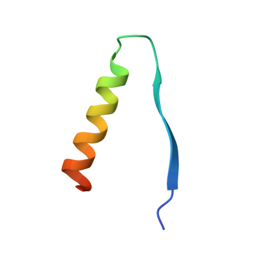
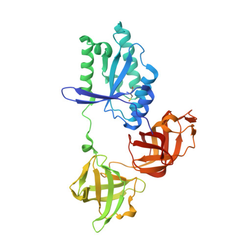
Molecular complementarity between tetracycline and the GTPase active site of elongation factor Tu.

Heffron, S.E.Mui, S.Aorora, A.Abel, K.Bergmann, E.Jurnak, F.

(2006) Acta Crystallogr D Biol Crystallogr 62: 1392-1400

  • DOI: https://doi.org/10.1107/S0907444906035426
  • Primary Citation Related Structures: 
    2HCJ, 2HDN

  • PubMed Abstract: 

    Two crystal forms of a complex between trypsin-modified elongation factor Tu-MgGDP from Escherichia coli and the antibiotic tetracycline have been solved by X-ray diffraction analysis to resolutions of 2.8 and 2.1 A, respectively. In the P2(1) form, cocrystals were grown from a solution mixture of the protein and tetracycline. Six copies of the trypsin-modified EF-Tu-MgGDP-tetracycline complex are arranged as three sets of dimers in the asymmetric unit. In the second crystal form, tetracycline was diffused into P4(3)2(1)2 crystals, resulting in a monomeric complex in the asymmetric unit. Atomic coordinates have been refined to crystallographic R factors of 18.0% for the P2(1) form and 20.0% for the P4(3)2(1)2 form. In both complexes, tetracycline makes significant interactions with the GTPase active site of EF-Tu. The phenoldiketone moiety of tetracycline interacts directly with the Mg(2+), the alpha-phosphate group of GDP and two amino acids, Thr25 and Asp80, which are conserved in the GX(4)GKS/T and DX(2)G sequence motifs found in all GTPases and many ATPases. The molecular complementarity, previously unrecognized between invariant groups present in all GTPase/ATPases and the active moiety of tetracycline, may have wide-ranging implications for all drugs containing the phenoldiketone moiety as well as for the design of new compounds targeted against a broad range of GTPases or ATPases.


  • Organizational Affiliation
    • Department of Physiology and Biophysics, 346-D Med Sci I, University of California, Irvine, CA 92697-4560, USA.

Macromolecule Content 

  • Total Structure Weight: 41.9 kDa 
  • Atom Count: 3,143 
  • Modeled Residue Count: 368 
  • Deposited Residue Count: 372 
  • Unique protein chains: 2

Macromolecules

Find similar proteins by:|  3D Structure
Entity ID: 1
MoleculeChains  Sequence LengthOrganismDetailsImage
Protein chain elongation factor EF-Tu37Escherichia coliMutation(s): 0 
EC: 3.6.5.3
UniProt
Find proteins for P0CE47 (Escherichia coli (strain K12))
Explore P0CE47 
Go to UniProtKB:  P0CE47
Entity Groups
Sequence Clusters30% Identity50% Identity70% Identity90% Identity95% Identity100% Identity
UniProt GroupP0CE47
Sequence Annotations
Expand
Reference Sequence
Find similar proteins by:|  3D Structure
Entity ID: 2
MoleculeChains  Sequence LengthOrganismDetailsImage
Protein chain elongation factor EF-Tu335Escherichia coliMutation(s): 2 
EC: 3.6.5.3
UniProt
Find proteins for Q1R5Y2 (Escherichia coli (strain UTI89 / UPEC))
Explore Q1R5Y2 
Go to UniProtKB:  Q1R5Y2
Entity Groups
Sequence Clusters30% Identity50% Identity70% Identity90% Identity95% Identity100% Identity
UniProt GroupQ1R5Y2
Sequence Annotations
Expand
Reference Sequence

Small Molecules

Ligands 6 Unique
IDChains Name / Formula / InChI Key2D Diagram3D Interactions
TAC

Query on TAC


Download:Ideal Coordinates CCD File
I [auth B]TETRACYCLINE
C22 H24 N2 O8
OFVLGDICTFRJMM-WESIUVDSSA-N
GDP

Query on GDP


Download:Ideal Coordinates CCD File
E [auth A]GUANOSINE-5'-DIPHOSPHATE
C10 H15 N5 O11 P2
QGWNDRXFNXRZMB-UUOKFMHZSA-N
SO4

Query on SO4


Download:Ideal Coordinates CCD File
D [auth A]SULFATE ION
O4 S
QAOWNCQODCNURD-UHFFFAOYSA-L
GLV

Query on GLV


Download:Ideal Coordinates CCD File
J [auth B]GLYOXYLIC ACID
C2 H2 O3
HHLFWLYXYJOTON-UHFFFAOYSA-N
MG

Query on MG


Download:Ideal Coordinates CCD File
C [auth A]MAGNESIUM ION
Mg
JLVVSXFLKOJNIY-UHFFFAOYSA-N
NA

Query on NA


Download:Ideal Coordinates CCD File
F [auth B],
G [auth B],
H [auth B]
SODIUM ION
Na
FKNQFGJONOIPTF-UHFFFAOYSA-N
Modified Residues  1 Unique
IDChains TypeFormula2D DiagramParent
CSO
Query on CSO
B
L-PEPTIDE LINKINGC3 H7 N O3 SCYS

Experimental Data & Validation

Experimental Data

  • Method: X-RAY DIFFRACTION
  • Resolution: 2.12 Å
  • R-Value Free:  0.234 (Depositor), 0.229 (DCC) 
  • R-Value Work:  0.200 (Depositor), 0.193 (DCC) 
Space Group: P 43 21 2
Unit Cell:
Length ( Å )Angle ( ˚ )
a = 69.11α = 90
b = 69.11β = 90
c = 157.33γ = 90
Software Package:
Software NamePurpose
CNSrefinement
EPMRphasing

Structure Validation

View Full Validation Report


Ligand Structure Quality Assessment 


Entry History 

Deposition Data

Revision History  (Full details and data files)

  • Version 1.0: 2006-10-31
    Type: Initial release
  • Version 1.1: 2008-05-01
    Changes: Version format compliance
  • Version 1.2: 2011-07-13
    Changes: Version format compliance
  • Version 1.3: 2018-09-05
    Changes: Data collection, Structure summary
  • Version 1.4: 2024-04-03
    Changes: Data collection, Database references, Derived calculations, Refinement description, Structure summary
  • Version 1.5: 2024-10-09
    Changes: Structure summary