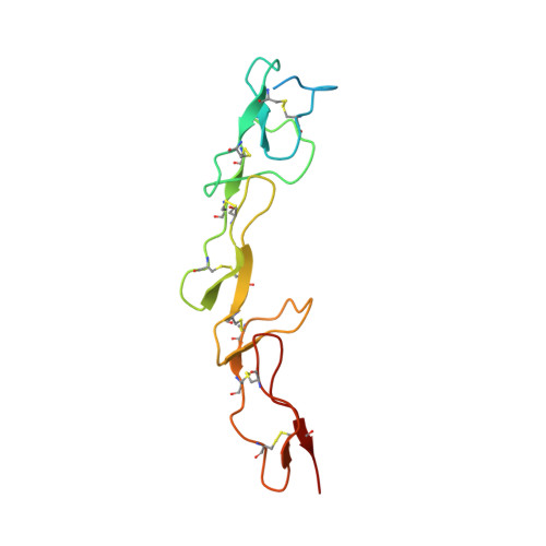
2H9G

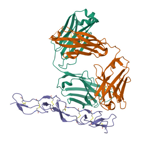
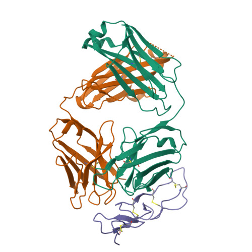
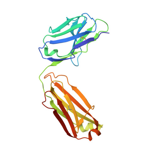
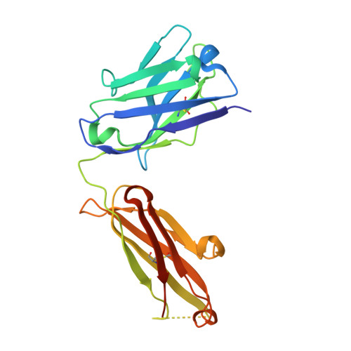
Crystal structure of phage derived Fab BdF1 with human Death Receptor 5 (DR5)


Experimental Data Snapshot

  • Method: X-RAY DIFFRACTION
  • Resolution: 2.32 Å
  • R-Value Free: 0.282 
  • R-Value Work: 0.228 
  • R-Value Observed: 0.234 

Starting Models: experimental
View more details

wwPDB Validation   3D Report Full Report


This is version 1.4 of the entry. See complete history


Literature

Activation of the Proapoptotic Death Receptor DR5 by Oligomeric Peptide and Antibody Agonists.

Li, B.Russell, S.J.Compaan, D.M.Totpal, K.Marsters, S.A.Ashkenazi, A.Cochran, A.G.Hymowitz, S.G.Sidhu, S.S.

(2006) J Mol Biol 361: 522-536

  • DOI: https://doi.org/10.1016/j.jmb.2006.06.042
  • Primary Citation of Related Structures:  
    2H9G

  • PubMed Abstract: 

    The cell-extrinsic apoptotic pathway triggers programmed cell death in response to certain ligands that bind to cell-surface death receptors. Apoptosis is essential for normal development and homeostasis in metazoans, and furthermore, selective activation of the cell-extrinsic pathway in tumor cells holds considerable promise for cancer therapy. We used phage display to identify peptides and synthetic antibodies that specifically bind to the human proapoptotic death receptor DR5. Despite great differences in overall size and structure, the DR5-binding peptides and antibodies shared a tripeptide motif, which was conserved within a disulfide-constrained loop of the peptides and the third complementarity determining region of the antibody heavy chains. The X-ray crystal structure of an antibody in complex with DR5 revealed that the tripeptide motif is buried at the core of the interface, confirming its central role in antigen recognition. We found that certain peptides and antibodies exhibited potent proapoptotic activity against DR5-expressing SK-MES-1 lung carcinoma cells. These phage-derived ligands may be useful for elucidating DR5 activation at the molecular level and for creating synthetic agonists of proapoptotic death receptors.


  • Organizational Affiliation

    Department of Protein Engineering, Genentech Inc., 1 DNA Way, South San Francisco, CA 94080, USA.


Macromolecules
Find similar proteins by:  (by identity cutoff)  |  3D Structure
Entity ID: 1
MoleculeChains Sequence LengthOrganismDetailsImage
Fab BdF1, light chainA,
D [auth L]
214Homo sapiensMutation(s): 0 
Entity Groups  
Sequence Clusters30% Identity50% Identity70% Identity90% Identity95% Identity100% Identity
Sequence Annotations
Expand
  • Reference Sequence
Find similar proteins by:  (by identity cutoff)  |  3D Structure
Entity ID: 2
MoleculeChains Sequence LengthOrganismDetailsImage
Fab BdF1, heavy chainB,
E [auth H]
228Homo sapiensMutation(s): 0 
Entity Groups  
Sequence Clusters30% Identity50% Identity70% Identity90% Identity95% Identity100% Identity
Sequence Annotations
Expand
  • Reference Sequence
Find similar proteins by:  (by identity cutoff)  |  3D Structure
Entity ID: 3
MoleculeChains Sequence LengthOrganismDetailsImage
Tumor necrosis factor receptor superfamily member 10B precursorC [auth R],
F [auth S]
130Homo sapiensMutation(s): 0 
Gene Names: TNFRSF10BDR5KILLERTRAILR2TRICK2ZTNFR9
UniProt & NIH Common Fund Data Resources
Find proteins for O14763 (Homo sapiens)
Explore O14763 
Go to UniProtKB:  O14763
PHAROS:  O14763
GTEx:  ENSG00000120889 
Entity Groups  
Sequence Clusters30% Identity50% Identity70% Identity90% Identity95% Identity100% Identity
UniProt GroupO14763
Sequence Annotations
Expand
  • Reference Sequence
Experimental Data & Validation

Experimental Data

  • Method: X-RAY DIFFRACTION
  • Resolution: 2.32 Å
  • R-Value Free: 0.282 
  • R-Value Work: 0.228 
  • R-Value Observed: 0.234 
  • Space Group: P 1 21 1
Unit Cell:
Length ( Å )Angle ( ˚ )
a = 99.814α = 90
b = 61.389β = 101.17
c = 108.269γ = 90
Software Package:
Software NamePurpose
REFMACrefinement
Blu-Icedata collection
HKL-2000data scaling
AMoREphasing

Structure Validation

View Full Validation Report


Entry History 

Deposition Data

Revision History  (Full details and data files)

  • Version 1.0: 2006-08-08
    Type: Initial release
  • Version 1.1: 2008-05-01
    Changes: Version format compliance
  • Version 1.2: 2011-07-13
    Changes: Advisory, Version format compliance
  • Version 1.3: 2023-08-30
    Changes: Data collection, Database references, Derived calculations, Refinement description
  • Version 1.4: 2024-11-20
    Changes: Structure summary