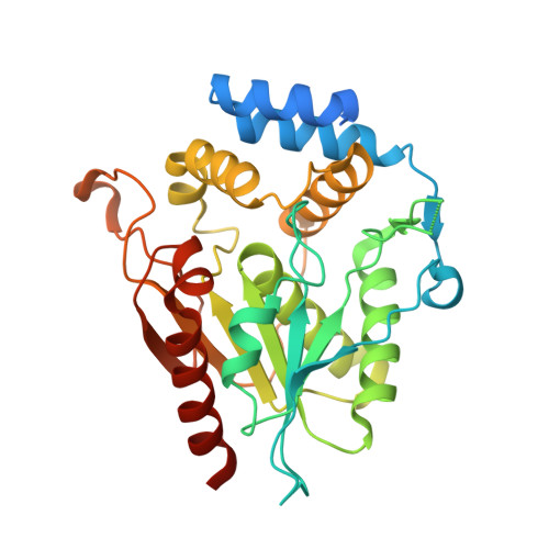
2H7Y

Pikromycin Thioesterase with covalent affinity label


Experimental Data Snapshot

  • Method: X-RAY DIFFRACTION
  • Resolution: 2.10 Å
  • R-Value Free: 0.236 
  • R-Value Work: 0.193 
  • R-Value Observed: 0.195 

Starting Model: experimental
View more details

wwPDB Validation   3D Report Full Report


Ligand Structure Quality Assessment 


This is version 1.4 of the entry. See complete history


Literature

Structural and Mechanistic Insights of Polyketide Macrolactonization from Polyketide-based Affinity Labels

Giraldes, J.W.Akey, D.L.Kittendorf, J.D.Sherman, D.H.Smith, J.S.Fecik, R.A.

(2006) Nat Chem Biol 2: 531-536

  • DOI: https://doi.org/10.1038/nchembio822
  • Primary Citation of Related Structures:  
    2H7X, 2H7Y

  • PubMed Abstract: 

    Polyketides are a diverse class of natural products having important clinical properties, including antibiotic, immunosuppressive and anticancer activities. They are biosynthesized by polyketide synthases (PKSs), which are modular, multienzyme complexes that sequentially condense simple carboxylic acid derivatives. The final reaction in many PKSs involves thioesterase-catalyzed cyclization of linear chain elongation intermediates. As the substrate in PKSs is presented by a tethered acyl carrier protein, introduction of substrate by diffusion is problematic, and no substrate-bound type I PKS domain structure has been reported so far. We describe the chemical synthesis of polyketide-based affinity labels that covalently modify the active site serine of excised pikromycin thioesterase from Streptomyces venezuelae. Crystal structures reported here of the affinity label-pikromycin thioesterase adducts provide important mechanistic insights. These results suggest that affinity labels can be valuable tools for understanding the mechanisms of individual steps within multifunctional PKSs and for directing rational engineering of PKS domains for combinatorial biosynthesis.


  • Organizational Affiliation

    Department of Medicinal Chemistry, University of Minnesota, 308 Harvard Street S.E., 8-101 Weaver-Densford Hall, Minneapolis, Minnesota 55455-0353, USA.


Macromolecules
Find similar proteins by:  (by identity cutoff)  |  3D Structure
Entity ID: 1
MoleculeChains Sequence LengthOrganismDetailsImage
Type I polyketide synthase PikAIV
A, B
319Streptomyces venezuelaeMutation(s): 0 
Gene Names: pikAIV
EC: 2.3.1.239 (UniProt), 2.3.1.240 (UniProt)
UniProt
Find proteins for Q9ZGI2 (Streptomyces venezuelae)
Explore Q9ZGI2 
Go to UniProtKB:  Q9ZGI2
Entity Groups  
Sequence Clusters30% Identity50% Identity70% Identity90% Identity95% Identity100% Identity
UniProt GroupQ9ZGI2
Sequence Annotations
Expand
  • Reference Sequence
Small Molecules
Ligands 4 Unique
IDChains Name / Formula / InChI Key2D Diagram3D Interactions
PSK
Query on PSK

Download Ideal Coordinates CCD File 
F [auth A],
J [auth B]
[(3R,4S)-4-HYDROXY-3-METHYL-2-OXOHEXYL]PHOSPHONIC ACID
C7 H15 O4 P
UXHVQAJQXZWLAW-RITPCOANSA-N
SO4
Query on SO4

Download Ideal Coordinates CCD File 
C [auth A],
D [auth A],
G [auth B]
SULFATE ION
O4 S
QAOWNCQODCNURD-UHFFFAOYSA-L
DMS
Query on DMS

Download Ideal Coordinates CCD File 
H [auth B],
I [auth B]
DIMETHYL SULFOXIDE
C2 H6 O S
IAZDPXIOMUYVGZ-UHFFFAOYSA-N
MG
Query on MG

Download Ideal Coordinates CCD File 
E [auth A]MAGNESIUM ION
Mg
JLVVSXFLKOJNIY-UHFFFAOYSA-N
Experimental Data & Validation

Experimental Data

  • Method: X-RAY DIFFRACTION
  • Resolution: 2.10 Å
  • R-Value Free: 0.236 
  • R-Value Work: 0.193 
  • R-Value Observed: 0.195 
  • Space Group: P 21 21 2
Unit Cell:
Length ( Å )Angle ( ˚ )
a = 107.736α = 90
b = 130.649β = 90
c = 56.176γ = 90
Software Package:
Software NamePurpose
REFMACrefinement
PDB_EXTRACTdata extraction
Blu-Icedata collection
SCALEPACKdata scaling
MOLREPphasing

Structure Validation

View Full Validation Report


Ligand Structure Quality Assessment 


Entry History 

Deposition Data

Revision History  (Full details and data files)

  • Version 1.0: 2006-09-19
    Type: Initial release
  • Version 1.1: 2008-05-01
    Changes: Version format compliance
  • Version 1.2: 2011-07-13
    Changes: Version format compliance
  • Version 1.3: 2023-08-30
    Changes: Data collection, Database references, Derived calculations, Refinement description
  • Version 1.4: 2024-11-06
    Changes: Structure summary