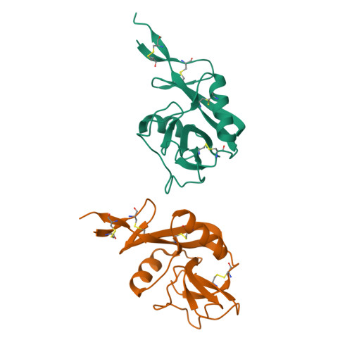
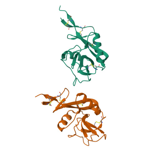
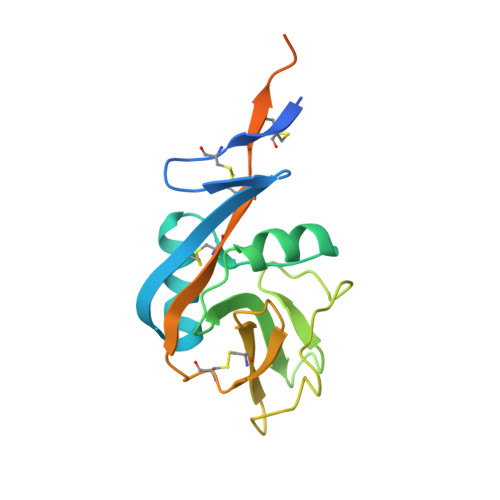
2H2R

Crystal structure of the human CD23 Lectin domain, apo form


Experimental Data Snapshot

  • Method: X-RAY DIFFRACTION
  • Resolution: 1.50 Å
  • R-Value Free: 0.165 
  • R-Value Work: 0.124 
  • R-Value Observed: 0.126 

Starting Model: experimental
View more details

wwPDB Validation   3D Report Full Report


This is version 1.6 of the entry. See complete history


Literature

Structural Changes in the Lectin Domain of CD23, the Low-Affinity IgE Receptor, upon Calcium Binding.

Wurzburg, B.A.Tarchevskaya, S.S.Jardetzky, T.S.

(2006) Structure 14: 1049-1058

  • DOI: https://doi.org/10.1016/j.str.2006.03.017
  • Primary Citation of Related Structures:  
    2H2R, 2H2T

  • PubMed Abstract: 

    CD23, the low-affinity receptor for IgE (Fc epsilonRII), regulates IgE synthesis and also mediates IgE-dependent antigen transport and processing. CD23 is a unique Fc receptor belonging to the C-type lectin-like domain superfamily and binds IgE in an unusual, non-lectin-like manner, requiring calcium but not carbohydrate. We have solved the high-resolution crystal structures of the human CD23 lectin domain in the presence and absence of Ca2+. The crystal structures differ significantly from a previously determined NMR structure and show that calcium binding occurs at the principal binding site, but not at an auxiliary site that appears to be absent in human CD23. Conformational differences between the apo and Ca2+ bound structures suggest how IgE-Fc binding can be both calcium-dependent and carbohydrate-independent.


  • Organizational Affiliation

    Department of Biochemistry, Molecular Biology and Cell Biology, Northwestern University, Evanston, Illinois 60208, USA.


Macromolecules
Find similar proteins by:  (by identity cutoff)  |  3D Structure
Entity ID: 1
MoleculeChains Sequence LengthOrganismDetailsImage
Low affinity immunoglobulin epsilon Fc receptor (Lymphocyte IgE receptor) (Fc-epsilon-RII)(Immunoglobulin E-binding factor) (CD23 antigen)
A, B
175Homo sapiensMutation(s): 2 
Gene Names: FCER2
UniProt & NIH Common Fund Data Resources
Find proteins for P06734 (Homo sapiens)
Explore P06734 
Go to UniProtKB:  P06734
PHAROS:  P06734
GTEx:  ENSG00000104921 
Entity Groups  
Sequence Clusters30% Identity50% Identity70% Identity90% Identity95% Identity100% Identity
UniProt GroupP06734
Sequence Annotations
Expand
  • Reference Sequence
Experimental Data & Validation

Experimental Data

  • Method: X-RAY DIFFRACTION
  • Resolution: 1.50 Å
  • R-Value Free: 0.165 
  • R-Value Work: 0.124 
  • R-Value Observed: 0.126 
  • Space Group: C 1 2 1
Unit Cell:
Length ( Å )Angle ( ˚ )
a = 112.354α = 90
b = 50.667β = 127.09
c = 75.061γ = 90
Software Package:
Software NamePurpose
REFMACrefinement
MAR345data collection
XDSdata scaling
CNSphasing

Structure Validation

View Full Validation Report


Entry History 

Deposition Data

Revision History  (Full details and data files)

  • Version 1.0: 2006-06-20
    Type: Initial release
  • Version 1.1: 2008-05-01
    Changes: Version format compliance
  • Version 1.2: 2011-07-13
    Changes: Version format compliance
  • Version 1.3: 2017-10-18
    Changes: Refinement description
  • Version 1.4: 2021-10-20
    Changes: Database references
  • Version 1.5: 2023-08-30
    Changes: Data collection, Refinement description
  • Version 1.6: 2024-11-20
    Changes: Structure summary