2GXA | pdb_00002gxa

Crystal structure of papillomavirus E1 hexameric helicase with ssDNA and MgADP


Experimental Data Snapshot

  • Method: X-RAY DIFFRACTION
  • Resolution: 3.15 Å
  • R-Value Free: 
    0.298 (Depositor) 
  • R-Value Work: 
    0.239 (Depositor), 0.214 (DCC) 
  • R-Value Observed: 
    0.239 (Depositor) 

Starting Model: experimental
View more details

wwPDB Validation 3D Report Full Report

Validation slider image for 2GXA

Ligand Structure Quality Assessment 


This is version 1.3 of the entry. See complete history

Literature

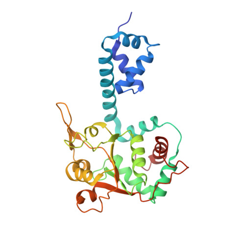
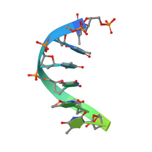
Mechanism of DNA translocation in a replicative hexameric helicase.

Enemark, E.J.Joshua-Tor, L.

(2006) Nature 442: 270-275

  • DOI: https://doi.org/10.1038/nature04943
  • Primary Citation Related Structures: 
    2GXA

  • PubMed Abstract: 

    The E1 protein of papillomavirus is a hexameric ring helicase belonging to the AAA + family. The mechanism that couples the ATP cycle to DNA translocation has been unclear. Here we present the crystal structure of the E1 hexamer with single-stranded DNA discretely bound within the hexamer channel and nucleotides at the subunit interfaces. This structure demonstrates that only one strand of DNA passes through the hexamer channel and that the DNA-binding hairpins of each subunit form a spiral 'staircase' that sequentially tracks the oligonucleotide backbone. Consecutively grouped ATP, ADP and apo configurations correlate with the height of the hairpin, suggesting a straightforward DNA translocation mechanism. Each subunit sequentially progresses through ATP, ADP and apo states while the associated DNA-binding hairpin travels from the top staircase position to the bottom, escorting one nucleotide of single-stranded DNA through the channel. These events permute sequentially around the ring from one subunit to the next.


  • Organizational Affiliation
    • W. M. Keck Structural Biology Laboratory, Cold Spring Harbor Laboratory, 1 Bungtown Road, Cold Spring Harbor, New York 11724, USA.

Macromolecule Content 

  • Total Structure Weight: 386.35 kDa 
  • Atom Count: 26,374 
  • Modeled Residue Count: 3,264 
  • Deposited Residue Count: 3,314 
  • Unique protein chains: 1
  • Unique nucleic acid chains: 1

Macromolecules


Find similar proteins by:|  3D Structure
Entity ID: 2
MoleculeChains  Sequence LengthOrganismDetailsImage
Replication protein E1274Deltapapillomavirus 4Mutation(s): 0 
Gene Names: E1
EC: 5.6.2.4
UniProt
Find proteins for P03116 (Bovine papillomavirus type 1)
Explore P03116 
Go to UniProtKB:  P03116
Entity Groups
Sequence Clusters30% Identity50% Identity70% Identity90% Identity95% Identity100% Identity
UniProt GroupP03116
Sequence Annotations
Expand
Reference Sequence
Find similar nucleic acids by:  Sequence
Entity ID: 1
MoleculeChains LengthOrganismImage
5'-D(*TP*TP*TP*TP*TP*TP*TP*TP*TP*TP*TP*TP*T)-3'A [auth M],
B [auth N]
13N/A
Sequence Annotations
Expand
Reference Sequence

Small Molecules

Ligands 3 Unique
IDChains Name / Formula / InChI Key2D Diagram3D Interactions
ADP

Query on ADP


Download:Ideal Coordinates CCD File
AA [auth F]
CA [auth G]
FA [auth H]
IA [auth J]
KA [auth K]
AA [auth F],
CA [auth G],
FA [auth H],
IA [auth J],
KA [auth K],
LA [auth L],
Q [auth A],
T [auth B],
V [auth C],
X [auth D],
Z [auth E]
ADENOSINE-5'-DIPHOSPHATE
C10 H15 N5 O10 P2
XTWYTFMLZFPYCI-KQYNXXCUSA-N
CL

Query on CL


Download:Ideal Coordinates CCD File
EA [auth H],
P [auth A],
S [auth B]
CHLORIDE ION
Cl
VEXZGXHMUGYJMC-UHFFFAOYSA-M
MG

Query on MG


Download:Ideal Coordinates CCD File
BA [auth G]
DA [auth H]
GA [auth I]
HA [auth J]
JA [auth K]
BA [auth G],
DA [auth H],
GA [auth I],
HA [auth J],
JA [auth K],
O [auth A],
R [auth B],
U [auth C],
W [auth D],
Y [auth E]
MAGNESIUM ION
Mg
JLVVSXFLKOJNIY-UHFFFAOYSA-N

Experimental Data & Validation

Experimental Data

  • Method: X-RAY DIFFRACTION
  • Resolution: 3.15 Å
  • R-Value Free:  0.298 (Depositor) 
  • R-Value Work:  0.239 (Depositor), 0.214 (DCC) 
  • R-Value Observed: 0.239 (Depositor) 
Space Group: P 1
Unit Cell:
Length ( Å )Angle ( ˚ )
a = 100.51α = 92.6
b = 100.881β = 111.46
c = 125.023γ = 106.01
Software Package:
Software NamePurpose
CNSrefinement
CBASSdata collection
HKL-2000data scaling
PHASERphasing

Structure Validation

View Full Validation Report


Ligand Structure Quality Assessment 


Entry History 

Deposition Data

Revision History  (Full details and data files)

  • Version 1.0: 2006-07-25
    Type: Initial release
  • Version 1.1: 2008-05-01
    Changes: Version format compliance
  • Version 1.2: 2011-07-13
    Changes: Version format compliance
  • Version 1.3: 2023-08-30
    Changes: Data collection, Database references, Derived calculations, Refinement description