2GRA

crystal structure of Human Pyrroline-5-carboxylate Reductase complexed with nadp


Experimental Data Snapshot

  • Method: X-RAY DIFFRACTION
  • Resolution: 3.10 Å
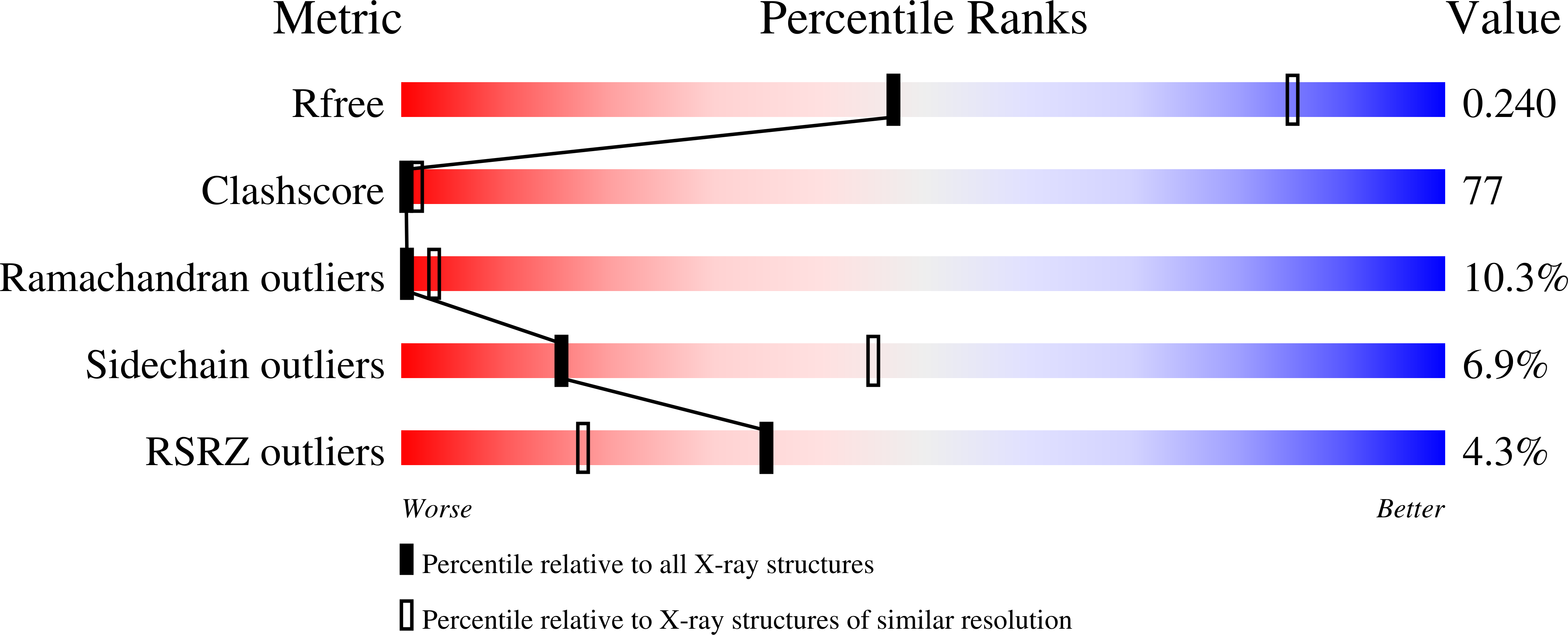
  • R-Value Free: 0.249 
  • R-Value Work: 0.228 
  • R-Value Observed: 0.230 

Starting Model: experimental
View more details

wwPDB Validation   3D Report Full Report


Ligand Structure Quality Assessment 


This is version 1.4 of the entry. See complete history


Literature

Crystal structure of human pyrroline-5-carboxylate reductase

Meng, Z.Lou, Z.Liu, Z.Li, M.Zhao, X.Bartlam, M.Rao, Z.

(2006) J Mol Biol 359: 1364-1377

  • DOI: https://doi.org/10.1016/j.jmb.2006.04.053
  • Primary Citation of Related Structures:  
    2GER, 2GR9, 2GRA

  • PubMed Abstract: 

    Pyrroline-5-carboxylate reductase (P5CR) is a universal housekeeping enzyme that catalyzes the reduction of Delta(1)-pyrroline-5-carboxylate (P5C) to proline using NAD(P)H as the cofactor. The enzymatic cycle between P5C and proline is very important for the regulation of amino acid metabolism, intracellular redox potential, and apoptosis. Here, we present the 2.8 Angstroms resolution structure of the P5CR apo enzyme, its 3.1 Angstroms resolution ternary complex with NAD(P)H and substrate-analog. The refined structures demonstrate a decameric architecture with five homodimer subunits and ten catalytic sites arranged around a peripheral circular groove. Mutagenesis and kinetic studies reveal the pivotal roles of the dinucleotide-binding Rossmann motif and residue Glu221 in the human enzyme. Human P5CR is thermostable and the crystals were grown at 37 degrees C. The enzyme is implicated in oxidation of the anti-tumor drug thioproline.


  • Organizational Affiliation

    Tsinghua-IBP Joint Research Group for Structural Biology, Tsinghua University, Beijing, China.


Macromolecules
Find similar proteins by:  (by identity cutoff)  |  3D Structure
Entity ID: 1
MoleculeChains Sequence LengthOrganismDetailsImage
Pyrroline-5-carboxylate reductase 1
A, B, C, D, E
277Homo sapiensMutation(s): 0 
EC: 1.5.1.2
UniProt & NIH Common Fund Data Resources
Find proteins for P32322 (Homo sapiens)
Explore P32322 
Go to UniProtKB:  P32322
PHAROS:  P32322
GTEx:  ENSG00000183010 
Entity Groups  
Sequence Clusters30% Identity50% Identity70% Identity90% Identity95% Identity100% Identity
UniProt GroupP32322
Sequence Annotations
Expand
  • Reference Sequence
Small Molecules
Ligands 2 Unique
IDChains Name / Formula / InChI Key2D Diagram3D Interactions
NAP
Query on NAP

Download Ideal Coordinates CCD File 
F [auth A],
H [auth B],
J [auth C],
L [auth D],
N [auth E]
NADP NICOTINAMIDE-ADENINE-DINUCLEOTIDE PHOSPHATE
C21 H28 N7 O17 P3
XJLXINKUBYWONI-NNYOXOHSSA-N
GLU
Query on GLU

Download Ideal Coordinates CCD File 
G [auth A],
I [auth B],
K [auth C],
M [auth D],
O [auth E]
GLUTAMIC ACID
C5 H9 N O4
WHUUTDBJXJRKMK-VKHMYHEASA-N
Experimental Data & Validation

Experimental Data

  • Method: X-RAY DIFFRACTION
  • Resolution: 3.10 Å
  • R-Value Free: 0.249 
  • R-Value Work: 0.228 
  • R-Value Observed: 0.230 
  • Space Group: C 1 2 1
Unit Cell:
Length ( Å )Angle ( ˚ )
a = 207.868α = 90
b = 123.502β = 121.97
c = 120.826γ = 90
Software Package:
Software NamePurpose
CrystalCleardata collection
HKL-2000data reduction
CNSrefinement
CrystalCleardata reduction
HKL-2000data scaling
CNSphasing

Structure Validation

View Full Validation Report


Ligand Structure Quality Assessment 


Entry History 

Deposition Data

Revision History  (Full details and data files)

  • Version 1.0: 2006-10-03
    Type: Initial release
  • Version 1.1: 2008-05-01
    Changes: Version format compliance
  • Version 1.2: 2011-07-13
    Changes: Derived calculations, Version format compliance
  • Version 1.3: 2023-10-25
    Changes: Data collection, Database references, Derived calculations, Refinement description
  • Version 1.4: 2024-12-25
    Changes: Advisory, Derived calculations, Structure summary