2GQ2 | pdb_00002gq2

Mycobacterium tuberculosis ThyX-NADP complex


Experimental Data Snapshot

  • Method: X-RAY DIFFRACTION
  • Resolution: 2.10 Å
  • R-Value Free: 
    0.255 (Depositor), 0.210 (DCC) 
  • R-Value Work: 
    0.212 (Depositor), 0.215 (DCC) 
  • R-Value Observed: 
    0.214 (Depositor) 

Starting Model: experimental
View more details

wwPDB Validation 3D Report Full Report

Validation slider image for 2GQ2

Ligand Structure Quality Assessment 


This is version 1.6 of the entry. See complete history

Literature

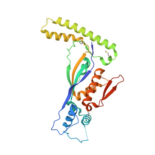
NADP+ expels both the co-factor and a substrate analog from the Mycobacterium tuberculosis ThyX active site: opportunities for anti-bacterial drug design.

Sampathkumar, P.Turley, S.Sibley, C.H.Hol, W.G.

(2006) J Mol Biology 360: 1-6

  • DOI: https://doi.org/10.1016/j.jmb.2006.04.061
  • Primary Citation Related Structures: 
    2GQ2

  • PubMed Abstract: 

    The novel flavin-dependent thymidylate synthase, ThyX, is absent in humans but several pathogenic bacteria depend exclusively on ThyX activity to synthesize thymidylate. Reduction of the enzyme-bound FAD by NADPH is suggested to be the critical first step in ThyX catalysis. We soaked Mycobacterium tuberculosis ThyX-FAD-BrdUMP ternary complex crystals in a solution containing NADP+ to gain structural insights into the reductive step of the catalytic cycle. Surprisingly, the NADP+ displaced both FAD and BrdUMP from the active site. In the resultant ThyX-NADP+ binary complex, the AMP moiety is bound in a deep pocket similar to that of the same moiety of FAD in the ternary complex, while the nicotinamide part of NADP+ is engaged in a limited number of contacts with ThyX. The additional 2'-phosphate group attached to the AMP ribose of NADP+ could be accommodated with minor rearrangement of water molecules. The newly introduced 2'-phosphate groups are engaged in water-mediated interactions across the non-crystallographic 2-fold axis of the ThyX tetramer, suggesting possibilities for design of high-affinity bivalent inhibitors of this intriguing enzyme.


  • Organizational Affiliation
    • Howard Hughes Medical Institute, University of Washington, Seattle, WA 98195, USA.

Macromolecule Content 

  • Total Structure Weight: 121.71 kDa 
  • Atom Count: 7,649 
  • Modeled Residue Count: 919 
  • Deposited Residue Count: 1,032 
  • Unique protein chains: 1

Macromolecules

Find similar proteins by:|  3D Structure
Entity ID: 1
MoleculeChains  Sequence LengthOrganismDetailsImage
Thymidylate synthase thyX
A, B, C, D
258Mycobacterium tuberculosis H37RvMutation(s): 5 
Gene Names: thyXRV2754C
EC: 2.1.1.148
UniProt
Find proteins for P9WG57 (Mycobacterium tuberculosis (strain ATCC 25618 / H37Rv))
Explore P9WG57 
Go to UniProtKB:  P9WG57
Entity Groups
Sequence Clusters30% Identity50% Identity70% Identity90% Identity95% Identity100% Identity
UniProt GroupP9WG57
Sequence Annotations
Expand
Reference Sequence

Small Molecules

Ligands 5 Unique
IDChains Name / Formula / InChI Key2D Diagram3D Interactions
NAP

Query on NAP


Download:Ideal Coordinates CCD File
FA [auth D],
J [auth A],
P [auth B],
W [auth C]
NADP NICOTINAMIDE-ADENINE-DINUCLEOTIDE PHOSPHATE
C21 H28 N7 O17 P3
XJLXINKUBYWONI-NNYOXOHSSA-N
PGE

Query on PGE


Download:Ideal Coordinates CCD File
K [auth A]TRIETHYLENE GLYCOL
C6 H14 O4
ZIBGPFATKBEMQZ-UHFFFAOYSA-N
IOD

Query on IOD


Download:Ideal Coordinates CCD File
AA [auth D]
BA [auth D]
CA [auth D]
DA [auth D]
E [auth A]
AA [auth D],
BA [auth D],
CA [auth D],
DA [auth D],
E [auth A],
F [auth A],
G [auth A],
H [auth A],
M [auth B],
N [auth B],
R [auth C],
S [auth C],
T [auth C],
U [auth C],
Z [auth D]
IODIDE ION
I
XMBWDFGMSWQBCA-UHFFFAOYSA-M
GOL

Query on GOL


Download:Ideal Coordinates CCD File
GA [auth D]
HA [auth D]
IA [auth D]
L [auth A]
Q [auth B]
GA [auth D],
HA [auth D],
IA [auth D],
L [auth A],
Q [auth B],
X [auth C],
Y [auth C]
GLYCEROL
C3 H8 O3
PEDCQBHIVMGVHV-UHFFFAOYSA-N
K

Query on K


Download:Ideal Coordinates CCD File
EA [auth D],
I [auth A],
O [auth B],
V [auth C]
POTASSIUM ION
K
NPYPAHLBTDXSSS-UHFFFAOYSA-N
Modified Residues  1 Unique
IDChains TypeFormula2D DiagramParent
MSE
Query on MSE
A, B, C, D
L-PEPTIDE LINKINGC5 H11 N O2 SeMET

Experimental Data & Validation

Experimental Data

  • Method: X-RAY DIFFRACTION
  • Resolution: 2.10 Å
  • R-Value Free:  0.255 (Depositor), 0.210 (DCC) 
  • R-Value Work:  0.212 (Depositor), 0.215 (DCC) 
  • R-Value Observed: 0.214 (Depositor) 
Space Group: P 1 21 1
Unit Cell:
Length ( Å )Angle ( ˚ )
a = 73.007α = 90
b = 78.09β = 95.67
c = 88.514γ = 90
Software Package:
Software NamePurpose
REFMACrefinement
ADSCdata collection
HKL-2000data scaling
AMoREphasing

Structure Validation

View Full Validation Report


Ligand Structure Quality Assessment 


Entry History 

Deposition Data

Revision History  (Full details and data files)

  • Version 1.0: 2006-06-27
    Type: Initial release
  • Version 1.1: 2008-05-01
    Changes: Version format compliance
  • Version 1.2: 2011-07-13
    Changes: Non-polymer description, Version format compliance
  • Version 1.3: 2021-10-20
    Changes: Database references, Derived calculations
  • Version 1.4: 2023-08-30
    Changes: Data collection, Refinement description
  • Version 1.5: 2023-11-15
    Changes: Data collection
  • Version 1.6: 2024-11-20
    Changes: Structure summary