2GO4 | pdb_00002go4

Crystal structure of Aquifex aeolicus LpxC complexed with TU-514


Experimental Data Snapshot

  • Method: X-RAY DIFFRACTION
  • Resolution: 2.70 Å
  • R-Value Free: 
    0.241 (Depositor), 0.224 (DCC) 
  • R-Value Work: 
    0.210 (Depositor), 0.215 (DCC) 

Starting Model: experimental
View more details

wwPDB Validation 3D Report Full Report

Validation slider image for 2GO4

Ligand Structure Quality Assessment 


This is version 1.4 of the entry. See complete history

Literature

Mechanistic Inferences from the Binding of Ligands to LpxC, a Metal-Dependent Deacetylase

Gennadios, H.A.Whittington, D.A.Li, X.Fierke, C.A.Christianson, D.W.

(2006) Biochemistry 45: 7940-7948

  • DOI: https://doi.org/10.1021/bi060823m
  • Primary Citation Related Structures: 
    2GO3, 2GO4

  • PubMed Abstract: 

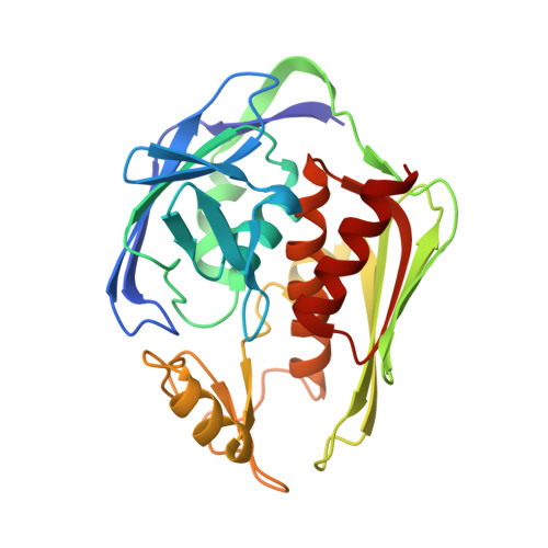
    The metal-dependent deacetylase LpxC catalyzes the first committed step of lipid A biosynthesis in Gram-negative bacteria. Accordingly, LpxC is an attractive target for the development of inhibitors that may serve as potential new antibiotics for the treatment of Gram-negative bacterial infections. Here, we report the 2.7 A resolution X-ray crystal structure of LpxC complexed with the substrate analogue inhibitor TU-514 and the 2.0 A resolution structure of LpxC complexed with imidazole. The X-ray crystal structure of LpxC complexed with TU-514 allows for a detailed examination of the coordination geometry of the catalytic zinc ion and other enzyme-inhibitor interactions in the active site. The hydroxamate group of TU-514 forms a bidentate chelate complex with the zinc ion and makes hydrogen bond interactions with conserved active site residues E78, H265, and T191. The inhibitor C-4 hydroxyl group makes direct hydrogen bond interactions with E197 and H58. Finally, the C-3 myristate moiety of the inhibitor binds in the hydrophobic tunnel of the active site. These intermolecular interactions provide a foundation for understanding structural aspects of enzyme-substrate and enzyme-inhibitor affinity. Comparison of the TU-514 complex with cacodylate and imidazole complexes suggests a possible substrate diphosphate binding site and highlights residues that may stabilize the tetrahedral intermediate and its flanking transition states in catalysis. Evidence of a catalytic zinc ion in the native zinc enzyme coordinated by H79, H238, D242, and two water molecules with square pyramidal geometry is also presented. These results suggest that the native state of this metallohydrolase may contain a pentacoordinate zinc ion, which contrasts with the native states of archetypical zinc hydrolases such as thermolysin and carboxypeptidase A.


  • Organizational Affiliation
    • Roy and Diana Vagelos Laboratories, Department of Chemistry, University of Pennsylvania, Philadelphia, Pennsylvania 19104-6323, USA.

Macromolecule Content 

  • Total Structure Weight: 62.2 kDa 
  • Atom Count: 4,420 
  • Modeled Residue Count: 532 
  • Deposited Residue Count: 534 
  • Unique protein chains: 1

Macromolecules

Find similar proteins by:|  3D Structure
Entity ID: 1
MoleculeChains  Sequence LengthOrganismDetailsImage
UDP-3-O-[3-hydroxymyristoyl] N-acetylglucosamine deacetylase
A, B
267Aquifex aeolicusMutation(s): 1 
Gene Names: lpxCenvA
EC: 3.5.1 (PDB Primary Data), 3.5.1.108 (UniProt)
UniProt
Find proteins for O67648 (Aquifex aeolicus (strain VF5))
Explore O67648 
Go to UniProtKB:  O67648
Entity Groups
Sequence Clusters30% Identity50% Identity70% Identity90% Identity95% Identity100% Identity
UniProt GroupO67648
Sequence Annotations
Expand
Reference Sequence

Small Molecules

Ligands 3 Unique
IDChains Name / Formula / InChI Key2D Diagram3D Interactions
TUX

Query on TUX


Download:Ideal Coordinates CCD File
G [auth A],
J [auth B]
1,5-ANHYDRO-2-C-(CARBOXYMETHYL-N-HYDROXYAMIDE)-2-DEOXY-3-O-MYRISTOYL-D-GLUCITOL
C22 H41 N O7
INAPDIIIYSWKOC-XHIHJMKYSA-N
ZN

Query on ZN


Download:Ideal Coordinates CCD File
C [auth A],
D [auth A],
E [auth A],
H [auth B],
I [auth B]
ZINC ION
Zn
PTFCDOFLOPIGGS-UHFFFAOYSA-N
CL

Query on CL


Download:Ideal Coordinates CCD File
F [auth A]CHLORIDE ION
Cl
VEXZGXHMUGYJMC-UHFFFAOYSA-M

Experimental Data & Validation

Experimental Data

  • Method: X-RAY DIFFRACTION
  • Resolution: 2.70 Å
  • R-Value Free:  0.241 (Depositor), 0.224 (DCC) 
  • R-Value Work:  0.210 (Depositor), 0.215 (DCC) 
Space Group: P 61
Unit Cell:
Length ( Å )Angle ( ˚ )
a = 100.729α = 90
b = 100.729β = 90
c = 121.299γ = 120
Software Package:
Software NamePurpose
HKL-2000data collection
HKL-2000data reduction
CNSrefinement
HKL-2000data scaling
CNSphasing

Structure Validation

View Full Validation Report


Ligand Structure Quality Assessment 


Entry History 

Deposition Data

Revision History  (Full details and data files)

  • Version 1.0: 2006-07-04
    Type: Initial release
  • Version 1.1: 2008-05-01
    Changes: Version format compliance
  • Version 1.2: 2011-07-13
    Changes: Derived calculations, Version format compliance
  • Version 1.3: 2021-10-20
    Changes: Database references, Derived calculations
  • Version 1.4: 2023-08-30
    Changes: Data collection, Refinement description