2GLO | pdb_00002glo

Solution structure of the Brinker DNA binding domain in complex with the omb enhancer


Experimental Data Snapshot

  • Method: SOLUTION NMR
  • Conformers Calculated: 100 
  • Conformers Submitted: 20 
  • Selection Criteria: structures with the lowest energy 

wwPDB Validation 3D Report Full Report

Validation slider image for 2GLO

This is version 1.4 of the entry. See complete history

Literature

DNA recognition by the brinker repressor - an extreme case of coupling between binding and folding

Cordier, F.Hartmann, B.Rogowski, M.Affolter, M.Grzesiek, S.

(2006) J Mol Biology 361: 659-672

  • DOI: https://doi.org/10.1016/j.jmb.2006.06.045
  • Primary Citation Related Structures: 
    2GLO

  • PubMed Abstract: 

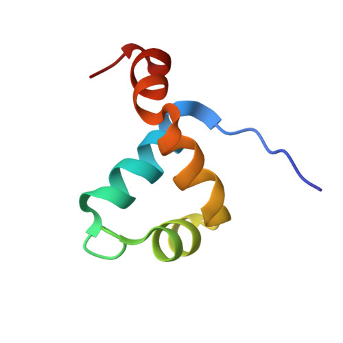
    The Brinker (Brk) nuclear repressor is a major element of the Drosophila Decapentaplegic morphogen signaling pathway. Its N-terminal part has weak homology to the Antennapedia homeodomain and binds to GC-rich DNA sequences. We have investigated the conformation and dynamics of the N-terminal 101 amino acid residues of Brk in the absence and in the presence of cognate DNA by solution NMR spectroscopy. In the absence of DNA, Brk is unfolded and highly flexible throughout the entire backbone. Addition of cognate DNA induces the formation of a well-folded structure for residues R46 to R95. This structure consists of four helices forming a helix-turn-helix motif that differs from homeodomains, but has similarities to the Tc3 transposase, the Pax-6 Paired domain, and the human centromere-binding protein. The GC-rich DNA recognition can be explained by specific major groove hydrogen bonds from the N-terminal end of helix alpha3. The transition from a highly flexible, completely unfolded conformation in the absence of DNA to a well-formed structure in the complex presents a very extreme case of the "coupling of binding and folding" phenomenon.


  • Organizational Affiliation
    • Division of Structural Biology, Biozentrum der Universität Basel, Klingelbergstrasse 70, CH-4056 Basel, Switzerland.

Macromolecule Content 

  • Total Structure Weight: 14.39 kDa 
  • Atom Count: 982 
  • Modeled Residue Count: 83 
  • Deposited Residue Count: 83 
  • Unique protein chains: 1
  • Unique nucleic acid chains: 2

Macromolecules


Find similar proteins by:|  3D Structure
Entity ID: 3
MoleculeChains  Sequence LengthOrganismDetailsImage
brinker CG9653-PAC [auth A]59Drosophila melanogasterMutation(s): 0 
Gene Names: brinker
UniProt
Find proteins for Q9XTN4 (Drosophila melanogaster)
Explore Q9XTN4 
Go to UniProtKB:  Q9XTN4
Entity Groups
Sequence Clusters30% Identity50% Identity70% Identity90% Identity95% Identity100% Identity
UniProt GroupQ9XTN4
Sequence Annotations
Expand
Reference Sequence
Find similar nucleic acids by:  Sequence
Entity ID: 1
MoleculeChains LengthOrganismImage
5'-D(*TP*GP*AP*GP*GP*CP*GP*TP*CP*AP*AP*C)-3'A [auth B]12N/A
Sequence Annotations
Expand
Reference Sequence
Find similar nucleic acids by:  Sequence
Entity ID: 2
MoleculeChains LengthOrganismImage
5'-D(*GP*TP*TP*GP*AP*CP*GP*CP*CP*TP*CP*A)-3'B [auth C]12N/A
Sequence Annotations
Expand
Reference Sequence

Experimental Data & Validation

Experimental Data

  • Method: SOLUTION NMR
  • Conformers Calculated: 100 
  • Conformers Submitted: 20 
  • Selection Criteria: structures with the lowest energy 

Structure Validation

View Full Validation Report


Entry History 

Deposition Data

Revision History  (Full details and data files)

  • Version 1.0: 2006-08-29
    Type: Initial release
  • Version 1.1: 2008-05-01
    Changes: Version format compliance
  • Version 1.2: 2011-07-13
    Changes: Version format compliance
  • Version 1.3: 2022-03-09
    Changes: Data collection, Database references, Derived calculations
  • Version 1.4: 2024-05-29
    Changes: Data collection