2GHV | pdb_00002ghv

Crystal structure of SARS spike protein receptor binding domain


Experimental Data Snapshot

  • Method: X-RAY DIFFRACTION
  • Resolution: 2.20 Å
  • R-Value Free: 
    0.213 (Depositor), 0.223 (DCC) 
  • R-Value Work: 
    0.182 (Depositor), 0.190 (DCC) 
  • R-Value Observed: 
    0.184 (Depositor) 

wwPDB Validation 3D Report Full Report

Validation slider image for 2GHV

This is version 1.3 of the entry. See complete history

Literature

Structural basis of neutralization by a human anti-severe acute respiratory syndrome spike protein antibody, 80R.

Hwang, W.C.Lin, Y.Santelli, E.Sui, J.Jaroszewski, L.Stec, B.Farzan, M.Marasco, W.A.Liddington, R.C.

(2006) J Biological Chem 281: 34610-34616

  • DOI: https://doi.org/10.1074/jbc.M603275200
  • Primary Citation Related Structures: 
    2GHV, 2GHW

  • PubMed Abstract: 

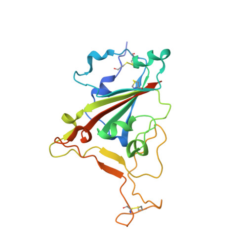
    Severe acute respiratory syndrome (SARS) is a newly emerged infectious disease that caused pandemic spread in 2003. The etiological agent of SARS is a novel coronavirus (SARS-CoV). The coronaviral surface spike protein S is a type I transmembrane glycoprotein that mediates initial host binding via the cell surface receptor angiotensin-converting enzyme 2 (ACE2), as well as the subsequent membrane fusion events required for cell entry. Here we report the crystal structure of the S1 receptor binding domain (RBD) in complex with a neutralizing antibody, 80R, at 2.3 A resolution, as well as the structure of the uncomplexed S1 RBD at 2.2 A resolution. We show that the 80R-binding epitope on the S1 RBD overlaps very closely with the ACE2-binding site, providing a rationale for the strong binding and broad neutralizing ability of the antibody. We provide a structural basis for the differential effects of certain mutations in the spike protein on 80R versus ACE2 binding, including escape mutants, which should facilitate the design of immunotherapeutics to treat a future SARS outbreak. We further show that the RBD of S1 forms dimers via an extensive interface that is disrupted in receptor- and antibody-bound crystal structures, and we propose a role for the dimer in virus stability and infectivity.


  • Organizational Affiliation
    • Infectious and Inflammatory Disease Center, Burnham Institute for Medical Research, La Jolla, California 92037, USA.

Macromolecule Content 

  • Total Structure Weight: 45.77 kDa 
  • Atom Count: 3,096 
  • Modeled Residue Count: 366 
  • Deposited Residue Count: 406 
  • Unique protein chains: 1

Macromolecules

Find similar proteins by:|  3D Structure
Entity ID: 1
MoleculeChains  Sequence LengthOrganismDetailsImage
Spike glycoproteinA [auth E],
B [auth C]
203Severe acute respiratory syndrome-related coronavirusMutation(s): 0 
Gene Names: S
UniProt
Find proteins for P59594 (Severe acute respiratory syndrome coronavirus)
Explore P59594 
Go to UniProtKB:  P59594
Entity Groups
Sequence Clusters30% Identity50% Identity70% Identity90% Identity95% Identity100% Identity
UniProt GroupP59594
Sequence Annotations
Expand
Reference Sequence

Experimental Data & Validation

Experimental Data

  • Method: X-RAY DIFFRACTION
  • Resolution: 2.20 Å
  • R-Value Free:  0.213 (Depositor), 0.223 (DCC) 
  • R-Value Work:  0.182 (Depositor), 0.190 (DCC) 
  • R-Value Observed: 0.184 (Depositor) 
Space Group: P 43 21 2
Unit Cell:
Length ( Å )Angle ( ˚ )
a = 75.884α = 90
b = 75.884β = 90
c = 235.863γ = 90
Software Package:
Software NamePurpose
REFMACrefinement
Blu-Icedata collection
HKL-2000data scaling
PHASERphasing

Structure Validation

View Full Validation Report


Entry History 

Deposition Data

Revision History  (Full details and data files)

  • Version 1.0: 2006-09-19
    Type: Initial release
  • Version 1.1: 2008-05-01
    Changes: Version format compliance
  • Version 1.2: 2011-07-13
    Changes: Advisory, Version format compliance
  • Version 1.3: 2024-11-20
    Changes: Data collection, Database references, Structure summary