2GH0 | pdb_00002gh0

Growth factor/receptor complex


Experimental Data Snapshot

  • Method: X-RAY DIFFRACTION
  • Resolution: 1.92 Å
  • R-Value Free: 
    0.263 (Depositor) 
  • R-Value Work: 
    0.219 (Depositor), 0.216 (DCC) 
  • R-Value Observed: 
    0.222 (Depositor) 

wwPDB Validation 3D Report Full Report

Validation slider image for 2GH0

Ligand Structure Quality Assessment 


This is version 2.1 of the entry. See complete history

Literature

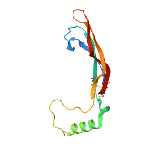
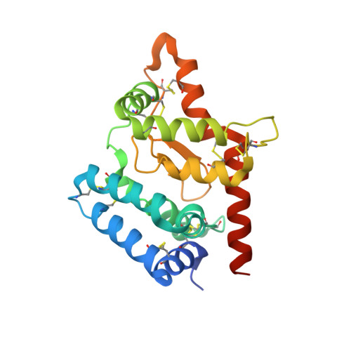
Structure of Artemin Complexed with Its Receptor GFRalpha3: Convergent Recognition of Glial Cell Line-Derived Neurotrophic Factors.

Wang, X.Baloh, R.H.Milbrandt, J.Garcia, K.C.

(2006) Structure 14: 1083-1092

  • DOI: https://doi.org/10.1016/j.str.2006.05.010
  • Primary Citation Related Structures: 
    2GH0, 2GYR, 2GYZ

  • PubMed Abstract: 

    Artemin (ARTN) is a member of the glial cell line-derived neurotrophic factor (GDNF) family ligands (GFLs) which regulate the development and maintenance of many neuronal populations in the mammalian nervous system. Here we report the 1.92 A crystal structure of the complex formed between ARTN and its receptor GFRalpha3, which is the initiating step in the formation of a ternary signaling complex containing the shared RET receptor. It represents a new receptor-ligand interaction mode for the TGF-beta superfamily that reveals both conserved and specificity-determining anchor points for all GFL-GFRalpha pairs. In tandem with the complex structure, cellular studies using receptor chimeras implicate dyad-symmetric composite interfaces for recruitment and dimerization of RET, leading to intracellular signaling. These studies should facilitate the functional dissection of the specific versus pleiotropic roles of this system in neurobiology, as well as its exploitation for therapeutic applications.


  • Organizational Affiliation
    • Howard Hughes Medical Institute, Stanford University School of Medicine, Department of Microbiology and Immunology, Stanford, California 94305-5124, USA.

Macromolecule Content 

  • Total Structure Weight: 70.6 kDa 
  • Atom Count: 4,881 
  • Modeled Residue Count: 598 
  • Deposited Residue Count: 628 
  • Unique protein chains: 2

Macromolecules

Find similar proteins by:|  3D Structure
Entity ID: 1
MoleculeChains  Sequence LengthOrganismDetailsImage
arteminA [auth C],
B [auth D]
101Homo sapiensMutation(s): 0 
UniProt & NIH Common Fund Data Resources
Find proteins for Q5T4W7 (Homo sapiens)
Explore Q5T4W7 
Go to UniProtKB:  Q5T4W7
PHAROS:  Q5T4W7
GTEx:  ENSG00000117407 
Entity Groups
Sequence Clusters30% Identity50% Identity70% Identity90% Identity95% Identity100% Identity
UniProt GroupQ5T4W7
Sequence Annotations
Expand
Reference Sequence
Find similar proteins by:|  3D Structure
Entity ID: 2
MoleculeChains  Sequence LengthOrganismDetailsImage
GDNF family receptor alpha-3C [auth A],
D [auth B]
213Homo sapiensMutation(s): 0 
Gene Names: GFRA3
UniProt & NIH Common Fund Data Resources
Find proteins for O60609 (Homo sapiens)
Explore O60609 
Go to UniProtKB:  O60609
PHAROS:  O60609
GTEx:  ENSG00000146013 
Entity Groups
Sequence Clusters30% Identity50% Identity70% Identity90% Identity95% Identity100% Identity
UniProt GroupO60609
Glycosylation
Glycosylation Sites: 1Go to GlyGen: O60609-1
Sequence Annotations
Expand
Reference Sequence

Oligosaccharides

Help  
Entity ID: 3
MoleculeChains Length2D Diagram GlycosylationD Interactions
alpha-D-mannopyranose-(1-3)-[alpha-D-mannopyranose-(1-6)]beta-D-mannopyranose-(1-4)-2-acetamido-2-deoxy-beta-D-glucopyranose-(1-4)-2-acetamido-2-deoxy-beta-D-glucopyranose
E
5N-Glycosylation
Glycosylation Resources
GlyTouCan: G22768VO
GlyCosmos: G22768VO
GlyGen: G22768VO

Small Molecules

Ligands 1 Unique
IDChains Name / Formula / InChI Key2D Diagram3D Interactions
NAG

Query on NAG


Download:Ideal Coordinates CCD File
F [auth B]2-acetamido-2-deoxy-beta-D-glucopyranose
C8 H15 N O6
OVRNDRQMDRJTHS-FMDGEEDCSA-N

Experimental Data & Validation

Experimental Data

  • Method: X-RAY DIFFRACTION
  • Resolution: 1.92 Å
  • R-Value Free:  0.263 (Depositor) 
  • R-Value Work:  0.219 (Depositor), 0.216 (DCC) 
  • R-Value Observed: 0.222 (Depositor) 
Space Group: P 1 21 1
Unit Cell:
Length ( Å )Angle ( ˚ )
a = 73.678α = 90
b = 41.46β = 103.56
c = 119.863γ = 90
Software Package:
Software NamePurpose
REFMACrefinement
ADSCdata collection
DENZOdata reduction
SCALEPACKdata scaling
PHASERphasing

Structure Validation

View Full Validation Report


Ligand Structure Quality Assessment 


Entry History 

Deposition Data

  • Released Date: 2006-06-27 
  • Deposition Author(s): Wang, X.Q.

Revision History  (Full details and data files)

  • Version 1.0: 2006-06-27
    Type: Initial release
  • Version 1.1: 2008-05-01
    Changes: Version format compliance
  • Version 1.2: 2011-07-13
    Changes: Advisory, Version format compliance
  • Version 2.0: 2020-07-29
    Type: Remediation
    Reason: Carbohydrate remediation
    Changes: Atomic model, Data collection, Database references, Derived calculations, Structure summary
  • Version 2.1: 2024-10-09
    Changes: Data collection, Database references, Structure summary