2GGF | pdb_00002ggf

Solution structure of the MA3 domain of human Programmed cell death 4


Experimental Data Snapshot

  • Method: SOLUTION NMR
  • Conformers Calculated: 100 
  • Conformers Submitted: 20 
  • Selection Criteria: target function 

wwPDB Validation 3D Report Full Report

Validation slider image for 2GGF

This is version 1.4 of the entry. See complete history

Literature

Solution structure of the MA3 domain of human Programmed cell death 4

Nagata, T.Muto, Y.Inoue, M.Kigawa, T.Terada, T.Shirouzu, M.Yokoyama, S.RIKEN Structural Genomics/Proteomics Initiative (RSGI)

To be published.

Macromolecule Content 

  • Total Structure Weight: 15.45 kDa 
  • Atom Count: 1,081 
  • Modeled Residue Count: 137 
  • Deposited Residue Count: 137 
  • Unique protein chains: 1

Macromolecules

Find similar proteins by:|  3D Structure
Entity ID: 1
MoleculeChains  Sequence LengthOrganismDetailsImage
Programmed cell death 4, isoform 1137Homo sapiensMutation(s): 0 
Gene Names: PDCD4
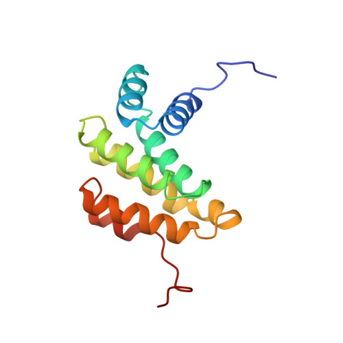
UniProt & NIH Common Fund Data Resources
Find proteins for Q53EL6 (Homo sapiens)
Explore Q53EL6 
Go to UniProtKB:  Q53EL6
PHAROS:  Q53EL6
GTEx:  ENSG00000150593 
Entity Groups
Sequence Clusters30% Identity50% Identity70% Identity90% Identity95% Identity100% Identity
UniProt GroupQ53EL6
Sequence Annotations
Expand
Reference Sequence

Experimental Data & Validation

Experimental Data

  • Method: SOLUTION NMR
  • Conformers Calculated: 100 
  • Conformers Submitted: 20 
  • Selection Criteria: target function 

Structure Validation

View Full Validation Report


Entry History 

Revision History  (Full details and data files)

  • Version 1.0: 2007-04-24
    Type: Initial release
  • Version 1.1: 2008-05-01
    Changes: Version format compliance
  • Version 1.2: 2011-07-13
    Changes: Version format compliance
  • Version 1.3: 2022-03-09
    Changes: Data collection, Database references, Derived calculations
  • Version 1.4: 2024-05-29
    Changes: Data collection