2GAZ | pdb_00002gaz

Mycobacterial lipoglycan presentation by CD1d


Experimental Data Snapshot

  • Method: X-RAY DIFFRACTION
  • Resolution: 2.61 Å
  • R-Value Free: 
    0.280 (Depositor), 0.277 (DCC) 
  • R-Value Work: 
    0.212 (Depositor), 0.211 (DCC) 
  • R-Value Observed: 
    0.216 (Depositor) 

Starting Model: experimental
View more details

wwPDB Validation 3D Report Full Report

Validation slider image for 2GAZ

Ligand Structure Quality Assessment 


This is version 2.3 of the entry. See complete history

Literature

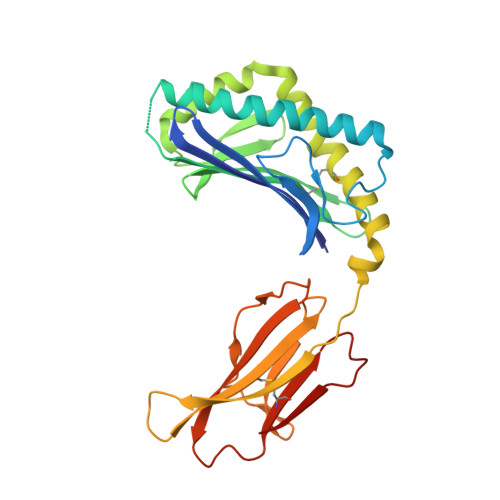
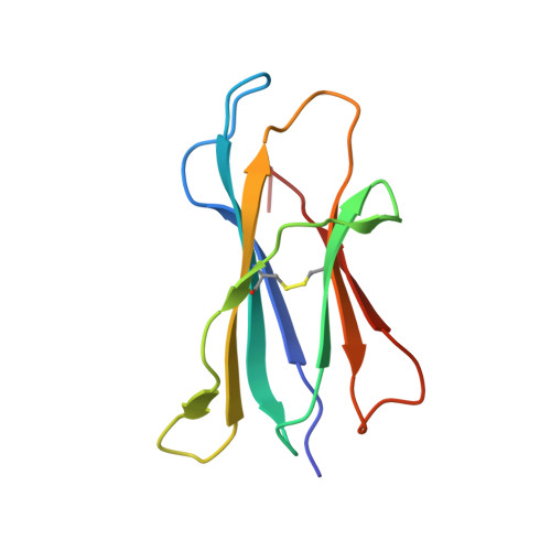
Structural characterization of mycobacterial phosphatidylinositol mannoside binding to mouse CD1d.

Zajonc, D.M.Ainge, G.D.Painter, G.F.Severn, W.B.Wilson, I.A.

(2006) J Immunol 177: 4577-4583

  • DOI: https://doi.org/10.4049/jimmunol.177.7.4577
  • Primary Citation Related Structures: 
    2GAZ

  • PubMed Abstract: 

    Mycobacterial phosphatidylinositol tetramannosides (PIM4) are agonists for a distinct population of invariant human (Valpha24) and mouse (Valpha14) NKT cells, when presented by CD1d. We determined the crystal structure at 2.6-A resolution of mouse CD1d bound to a synthetic dipalmitoyl-PIM2. Natural PIM2, which differs in its fatty acid composition is a biosynthetic precursor of PIM4, PIM6, lipomannan, and lipoarabinomannan. The PIM2 headgroup (inositol-dimannoside) is the most complex to date among all the crystallized CD1d ligands and is remarkably ordered in the CD1d binding groove. A specific hydrogen-bonding network between PIM2 and CD1d orients the headgroup in the center of the binding groove and above the A' pocket. A central cluster of hydrophilic CD1d residues (Asp(153), Thr(156), Ser(76), Arg(79)) interacts with the phosphate, inositol, and alpha1-alpha6-linked mannose of the headgroup, whereas additional specificity for the alpha1- and alpha2-linked mannose is conferred by Thr(159). The additional two mannoses in PIM4, relative to PIM2, are located at the distal 6' carbon of the alpha1-alpha6-linked mannose and would project away from the CD1d binding groove for interaction with the TCR. Compared with other CD1d-sphingolipid structures, PIM2 has an increased number of polar interactions between its headgroup and CD1, but reduced specificity for the diacylglycerol backbone. Thus, novel NKT cell agonists can be designed that focus on substitutions of the headgroup rather than on reducing lipid chain length, as in OCH and PBS-25, two potent variants of the highly stimulatory invariant NKT cell agonist alpha-galactosylceramide.


  • Organizational Affiliation
    • Department of Molecular Biology, The Scripps Research Institute, 10550 North Torrey Pines Road, La Jolla, CA 92037, USA.

Macromolecule Content 

  • Total Structure Weight: 46.78 kDa 
  • Atom Count: 3,182 
  • Modeled Residue Count: 367 
  • Deposited Residue Count: 384 
  • Unique protein chains: 2

Macromolecules

Find similar proteins by:|  3D Structure
Entity ID: 1
MoleculeChains  Sequence LengthOrganismDetailsImage
T-cell surface glycoprotein CD1d1285Mus musculusMutation(s): 0 
Gene Names: Cd1d1Cd1.1
UniProt & NIH Common Fund Data Resources
Find proteins for P11609 (Mus musculus)
Explore P11609 
Go to UniProtKB:  P11609
IMPC:  MGI:107674
Entity Groups
Sequence Clusters30% Identity50% Identity70% Identity90% Identity95% Identity100% Identity
UniProt GroupP11609
Glycosylation
Glycosylation Sites: 3Go to GlyGen: P11609-1
Sequence Annotations
Expand
Reference Sequence
Find similar proteins by:|  3D Structure
Entity ID: 2
MoleculeChains  Sequence LengthOrganismDetailsImage
beta-2-microglobulin99Mus musculusMutation(s): 0 
Gene Names: B2m
UniProt & NIH Common Fund Data Resources
Find proteins for P01887 (Mus musculus)
Explore P01887 
Go to UniProtKB:  P01887
IMPC:  MGI:88127
Entity Groups
Sequence Clusters30% Identity50% Identity70% Identity90% Identity95% Identity100% Identity
UniProt GroupP01887
Sequence Annotations
Expand
Reference Sequence

Oligosaccharides

Help  
Entity ID: 3
MoleculeChains Length2D Diagram GlycosylationD Interactions
alpha-D-mannopyranose-(1-3)-[alpha-D-mannopyranose-(1-6)]beta-D-mannopyranose-(1-4)-2-acetamido-2-deoxy-beta-D-glucopyranose-(1-4)-2-acetamido-2-deoxy-beta-D-glucopyranose
C
5N-Glycosylation
Glycosylation Resources
GlyTouCan: G22768VO
GlyCosmos: G22768VO
GlyGen: G22768VO

Small Molecules

Ligands 2 Unique
IDChains Name / Formula / InChI Key2D Diagram3D Interactions
XPX

Query on XPX


Download:Ideal Coordinates CCD File
F [auth A](2R)-3-[(HYDROXY{[(2R,3R,5S,6R)-3,4,5-TRIHYDROXY-2,6-BIS(ALPHA-D-MANNOPYRANOSYLOXY)CYCLOHEXYL]OXY}PHOSPHORYL)OXY]PROPAN E-1,2-DIYL DIHEXADECANOATE
C53 H99 O23 P
XGAHMXUNNQXCDP-FKPQOHGQSA-N
NAG

Query on NAG


Download:Ideal Coordinates CCD File
D [auth A],
E [auth A]
2-acetamido-2-deoxy-beta-D-glucopyranose
C8 H15 N O6
OVRNDRQMDRJTHS-FMDGEEDCSA-N

Experimental Data & Validation

Experimental Data

  • Method: X-RAY DIFFRACTION
  • Resolution: 2.61 Å
  • R-Value Free:  0.280 (Depositor), 0.277 (DCC) 
  • R-Value Work:  0.212 (Depositor), 0.211 (DCC) 
  • R-Value Observed: 0.216 (Depositor) 
Space Group: P 21 21 21
Unit Cell:
Length ( Å )Angle ( ˚ )
a = 41.835α = 90
b = 110.757β = 90
c = 107.396γ = 90
Software Package:
Software NamePurpose
REFMACrefinement
HKL-2000data reduction
CCP4data scaling
MOLREPphasing

Structure Validation

View Full Validation Report


Ligand Structure Quality Assessment 


Entry History 

Deposition Data

  • Released Date: 2006-09-26 
  • Deposition Author(s): Zajonc, D.M.

Revision History  (Full details and data files)

  • Version 1.0: 2006-09-26
    Type: Initial release
  • Version 1.1: 2008-05-01
    Changes: Version format compliance
  • Version 1.2: 2011-07-13
    Changes: Advisory, Version format compliance
  • Version 2.0: 2020-07-29
    Type: Remediation
    Reason: Carbohydrate remediation
    Changes: Advisory, Atomic model, Data collection, Database references, Derived calculations, Source and taxonomy, Structure summary
  • Version 2.1: 2023-08-30
    Changes: Advisory, Data collection, Database references, Refinement description, Structure summary
  • Version 2.2: 2024-03-13
    Changes: Source and taxonomy, Structure summary
  • Version 2.3: 2024-10-30
    Changes: Structure summary