2G9E | pdb_00002g9e

Protonation-mediated structural flexibility in the F conjugation regulatory protein, TRAM


Experimental Data Snapshot

  • Method: X-RAY DIFFRACTION
  • Resolution: 1.80 Å
  • R-Value Free: 
    0.245 (Depositor), 0.244 (DCC) 
  • R-Value Work: 
    0.182 (Depositor), 0.181 (DCC) 
  • R-Value Observed: 
    0.185 (Depositor) 

Starting Model: experimental
View more details

wwPDB Validation 3D Report Full Report

Validation slider image for 2G9E

This is version 1.4 of the entry. See complete history

Literature

Protonation-mediated structural flexibility in the F conjugation regulatory protein, TraM.

Lu, J.Edwards, R.A.Wong, J.J.Manchak, J.Scott, P.G.Frost, L.S.Glover, J.N.

(2006) EMBO J 25: 2930-2939

  • DOI: https://doi.org/10.1038/sj.emboj.7601151
  • Primary Citation Related Structures: 
    2G7O, 2G9E

  • PubMed Abstract: 

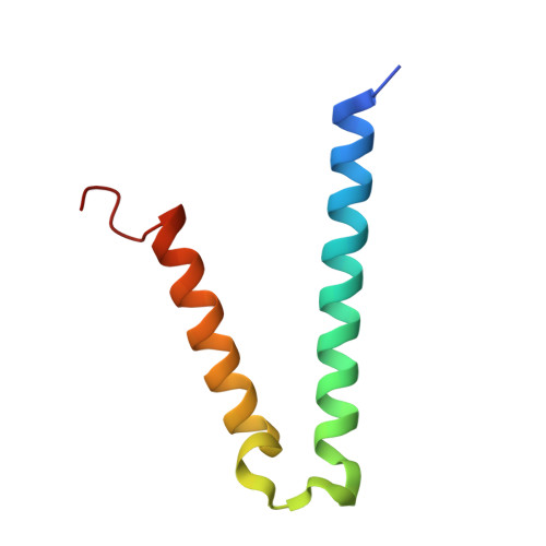
    TraM is essential for F plasmid-mediated bacterial conjugation, where it binds to the plasmid DNA near the origin of transfer, and recognizes a component of the transmembrane DNA transfer complex, TraD. Here we report the 1.40 A crystal structure of the TraM core tetramer (TraM58-127). TraM58-127 is a compact eight-helical bundle, in which the N-terminal helices from each protomer interact to form a central, parallel four-stranded coiled-coil, whereas each C-terminal helix packs in an antiparallel arrangement around the outside of the structure. Four protonated glutamic acid residues (Glu88) are packed in a hydrogen-bonded arrangement within the central four-helix bundle. Mutational and biophysical analyses indicate that this protonated state is in equilibrium with a deprotonated tetrameric form characterized by a lower helical content at physiological pH and temperature. Comparison of TraM to its Glu88 mutants predicted to stabilize the helical structure suggests that the protonated state is the active form for binding TraD in conjugation.


  • Organizational Affiliation
    • Department of Biochemistry, University of Alberta, Edmonton, Alberta, Canada.

Macromolecule Content 

  • Total Structure Weight: 7.96 kDa 
  • Atom Count: 585 
  • Modeled Residue Count: 68 
  • Deposited Residue Count: 70 
  • Unique protein chains: 1

Macromolecules

Find similar proteins by:|  3D Structure
Entity ID: 1
MoleculeChains  Sequence LengthOrganismDetailsImage
Protein traM70Escherichia coliMutation(s): 1 
Gene Names: traM
UniProt
Find proteins for P10026 (Escherichia coli (strain K12))
Explore P10026 
Go to UniProtKB:  P10026
Entity Groups
Sequence Clusters30% Identity50% Identity70% Identity90% Identity95% Identity100% Identity
UniProt GroupP10026
Sequence Annotations
Expand
Reference Sequence

Experimental Data & Validation

Experimental Data

  • Method: X-RAY DIFFRACTION
  • Resolution: 1.80 Å
  • R-Value Free:  0.245 (Depositor), 0.244 (DCC) 
  • R-Value Work:  0.182 (Depositor), 0.181 (DCC) 
  • R-Value Observed: 0.185 (Depositor) 
Space Group: I 4
Unit Cell:
Length ( Å )Angle ( ˚ )
a = 50.939α = 90
b = 50.939β = 90
c = 46.551γ = 90
Software Package:
Software NamePurpose
REFMACrefinement
DENZOdata reduction
SCALEPACKdata scaling
MOLREPphasing

Structure Validation

View Full Validation Report


Entry History 

Deposition Data

Revision History  (Full details and data files)

  • Version 1.0: 2006-06-13
    Type: Initial release
  • Version 1.1: 2008-05-01
    Changes: Version format compliance
  • Version 1.2: 2011-07-13
    Changes: Advisory, Derived calculations, Version format compliance
  • Version 1.3: 2021-10-20
    Changes: Database references
  • Version 1.4: 2023-08-30
    Changes: Data collection, Refinement description