2G9C | pdb_00002g9c

Modified pyrimidines Specifically bind the purine riboswitch


Experimental Data Snapshot

  • Method: X-RAY DIFFRACTION
  • Resolution: 1.70 Å
  • R-Value Free: 
    0.242 (Depositor), 0.235 (DCC) 
  • R-Value Work: 
    0.195 (Depositor), 0.187 (DCC) 
  • R-Value Observed: 
    0.231 (Depositor) 

wwPDB Validation 3D Report Full Report

Validation slider image for 2G9C

This is version 1.4 of the entry. See complete history

Literature

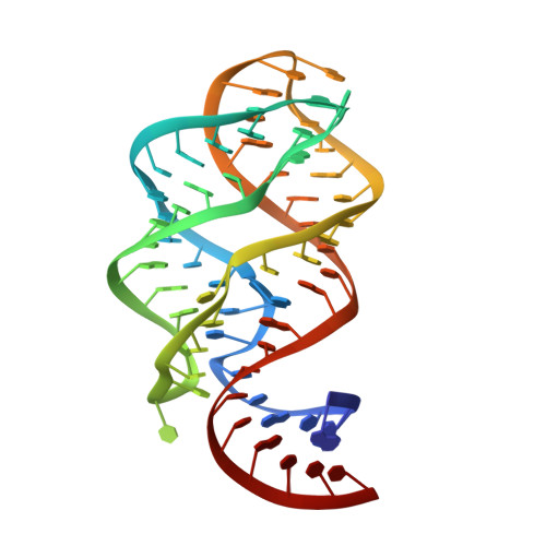
Modified pyrimidines specifically bind the purine riboswitch.

Gilbert, S.D.Mediatore, S.J.Batey, R.T.

(2006) J Am Chem Soc 128: 14214-14215

  • DOI: https://doi.org/10.1021/ja063645t
  • Primary Citation Related Structures: 
    2G9C

  • PubMed Abstract: 

    The purine riboswitch is a genetic regulatory element found in the 5'-untranslated regions of Gram-positive bacteria that regulates expression of the mRNA specifically in response to either guanine or adenine. We report that the adenine-responsive RNA element is also capable of specifically recognizing pyrimidine compounds bearing modifications at the 6- or 5,6-positions in a fashion similar to that of purine compounds. Using isothermal titration calorimetry and X-ray crystallography, the binding of these compounds is characterized.


  • Organizational Affiliation
    • Department of Chemistry and Biochemistry, University of Colorado at Boulder, Campus Box 215, Boulder, Colorado 80309-0215, USA.

Macromolecule Content 

  • Total Structure Weight: 23.63 kDa 
  • Atom Count: 1,872 
  • Modeled Residue Count: 67 
  • Deposited Residue Count: 67 
  • Unique nucleic acid chains: 1

Macromolecules

Find similar nucleic acids by:  (by identity cutoff) 
Entity ID: 1
MoleculeChains LengthOrganismImage
guanine riboswitch67N/A
Sequence Annotations
Expand
Reference Sequence

Small Molecules

Ligands 3 Unique
IDChains Name / Formula / InChI Key2D Diagram3D Interactions
NCO

Query on NCO


Download:Ideal Coordinates CCD File
C [auth A]
D [auth A]
E [auth A]
F [auth A]
G [auth A]
C [auth A],
D [auth A],
E [auth A],
F [auth A],
G [auth A],
H [auth A],
I [auth A],
J [auth A],
K [auth A],
L [auth A],
M [auth A],
N [auth A]
COBALT HEXAMMINE(III)
Co H18 N6
DYLMFCCYOUSRTK-UHFFFAOYSA-N
3AY

Query on 3AY


Download:Ideal Coordinates CCD File
O [auth A]PYRIMIDINE-2,4,6-TRIAMINE
C4 H7 N5
JTTIOYHBNXDJOD-UHFFFAOYSA-N
ACT

Query on ACT


Download:Ideal Coordinates CCD File
B [auth A]ACETATE ION
C2 H3 O2
QTBSBXVTEAMEQO-UHFFFAOYSA-M

Experimental Data & Validation

Experimental Data

  • Method: X-RAY DIFFRACTION
  • Resolution: 1.70 Å
  • R-Value Free:  0.242 (Depositor), 0.235 (DCC) 
  • R-Value Work:  0.195 (Depositor), 0.187 (DCC) 
  • R-Value Observed: 0.231 (Depositor) 
Space Group: C 1 2 1
Unit Cell:
Length ( Å )Angle ( ˚ )
a = 131.99α = 90
b = 35.1β = 90.43
c = 41.8γ = 90
Software Package:
Software NamePurpose
CNSrefinement
BOSdata collection
d*TREKdata scaling
CNSphasing

Structure Validation

View Full Validation Report


Entry History 

Deposition Data

Revision History  (Full details and data files)

  • Version 1.0: 2006-11-21
    Type: Initial release
  • Version 1.1: 2008-05-01
    Changes: Version format compliance
  • Version 1.2: 2011-07-13
    Changes: Version format compliance
  • Version 1.3: 2017-10-18
    Changes: Refinement description
  • Version 1.4: 2024-02-14
    Changes: Data collection, Database references, Derived calculations