2G8Q | pdb_00002g8q

The crystal structure of RNase A from monoclinic crystals at 100 K


Experimental Data Snapshot

  • Method: X-RAY DIFFRACTION
  • Resolution: 1.50 Å
  • R-Value Free: 
    0.218 (Depositor), 0.314 (DCC) 
  • R-Value Work: 
    0.185 (Depositor) 
  • R-Value Observed: 
    0.187 (Depositor) 

Starting Model: experimental
View more details

wwPDB Validation 3D Report Full Report

Validation slider image for 2G8Q

This is version 1.4 of the entry. See complete history

Literature

The binding of 3'-N-piperidine-4-carboxyl-3'-deoxy-ara-uridine to ribonuclease A in the crystal.

Leonidas, D.D.Maiti, T.K.Samanta, A.Dasgupta, S.Pathak, T.Zographos, S.E.Oikonomakos, N.G.

(2006) Bioorg Med Chem 14: 6055-6064

  • DOI: https://doi.org/10.1016/j.bmc.2006.05.011
  • Primary Citation Related Structures: 
    2G8Q, 2G8R

  • PubMed Abstract: 

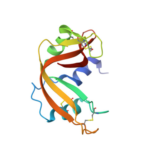
    The binding of a moderate inhibitor, 3'-N-piperidine-4-carboxyl-3'-deoxy-ara-uridine, to ribonuclease A has been studied by X-ray crystallography at 1.7A resolution. Two inhibitor molecules are bound in the central RNA binding cavity of RNase A exploiting interactions with residues from peripheral binding sites rather than from the active site of the enzyme. The uracyl moiety of the first inhibitor molecule occupies the purine-preferring site of RNase A, while the rest of the molecule projects to the solvent. The second inhibitor molecule binds with the carboxyl group at the pyrimidine recognition site and the uridine moiety exploits interactions with RNase A residues Lys66, His119 and Asp121. Comparative structural analysis of the 3'-N-piperidine-4-carboxyl-3'-deoxy-ara-uridine complex with other RNase A-ligand complexes provides a structural explanation of its potency. The crystal structure of the RNase A-3'-N-piperidine-4-carboxyl-3'-deoxy-ara-uridine complex provides evidence of a novel ligand-binding pattern in RNase A for 3'-N-aminonucleosides that was not anticipated by modelling studies, while it also suggests ways to improve the efficiency and selectivity of such compounds to develop pharmaceuticals against pathologies associated with RNase A homologues.


  • Organizational Affiliation
    • Institute of Organic and Pharmaceutical Chemistry, The National Hellenic Research Foundation, 48 Vas. Constantinou Avenue, 11635 Athens, Greece. ddl@eie.gr

Macromolecule Content 

  • Total Structure Weight: 27.42 kDa 
  • Atom Count: 2,286 
  • Modeled Residue Count: 248 
  • Deposited Residue Count: 248 
  • Unique protein chains: 1

Macromolecules

Find similar proteins by:|  3D Structure
Entity ID: 1
MoleculeChains  Sequence LengthOrganismDetailsImage
Ribonuclease pancreatic
A, B
124Bos taurusMutation(s): 0 
EC: 3.1.27.5 (PDB Primary Data), 4.6.1.18 (UniProt)
UniProt
Find proteins for P61823 (Bos taurus)
Explore P61823 
Go to UniProtKB:  P61823
Entity Groups
Sequence Clusters30% Identity50% Identity70% Identity90% Identity95% Identity100% Identity
UniProt GroupP61823
Sequence Annotations
Expand
Reference Sequence

Experimental Data & Validation

Experimental Data

  • Method: X-RAY DIFFRACTION
  • Resolution: 1.50 Å
  • R-Value Free:  0.218 (Depositor), 0.314 (DCC) 
  • R-Value Work:  0.185 (Depositor) 
  • R-Value Observed: 0.187 (Depositor) 
Space Group: C 1 2 1
Unit Cell:
Length ( Å )Angle ( ˚ )
a = 99.983α = 90
b = 32.551β = 90.48
c = 72.355γ = 90
Software Package:
Software NamePurpose
REFMACrefinement
DENZOdata reduction
SCALEPACKdata scaling

Structure Validation

View Full Validation Report


Entry History 

Deposition Data

Revision History  (Full details and data files)

  • Version 1.0: 2006-08-15
    Type: Initial release
  • Version 1.1: 2008-05-01
    Changes: Version format compliance
  • Version 1.2: 2011-07-13
    Changes: Advisory, Version format compliance
  • Version 1.3: 2023-08-30
    Changes: Data collection, Database references, Refinement description
  • Version 1.4: 2024-10-30
    Changes: Structure summary