2G81 | pdb_00002g81

Crystal Structure of the Bowman-Birk Inhibitor from Vigna unguiculata Seeds in Complex with Beta-trypsin at 1.55 Angstrons Resolution


Experimental Data Snapshot

  • Method: X-RAY DIFFRACTION
  • Resolution: 1.55 Å
  • R-Value Free: 
    0.169 (Depositor) 
  • R-Value Work: 
    0.154 (Depositor) 
  • R-Value Observed: 
    0.155 (Depositor) 

Starting Model: experimental
View more details

wwPDB Validation 3D Report Full Report

Validation slider image for 2G81

This is version 1.4 of the entry. See complete history

Literature

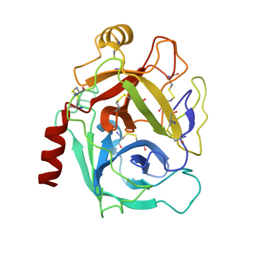
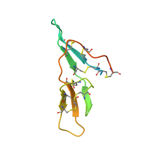
Crystal Structure of the Bowman-Birk Inhibitor from Vigna unguiculata Seeds in Complex with {beta}-Trypsin at 1.55 A Resolution and Its Structural Properties in Association with Proteinases

Barbosa, J.A.R.G.Silva, L.P.Teles, R.C.L.Esteves, G.F.Azevedo, R.B.Ventura, M.M.Freitas, S.M.

(2007) Biophys J 92: 1638-1650

  • DOI: https://doi.org/10.1529/biophysj.106.090555
  • Primary Citation Related Structures: 
    2G81

  • PubMed Abstract: 

    The structure of the Bowman-Birk inhibitor from Vigna unguiculata seeds (BTCI) in complex with beta-trypsin was solved and refined at 1.55 A to a crystallographic R(factor) of 0.154 and R(free) of 0.169, and represents the highest resolution for a Bowman-Birk inhibitor structure to date. The BTCI-trypsin interface is stabilized by hydrophobic contacts and hydrogen bonds, involving two waters and a polyethylene glycol molecule. The conformational rigidity of the reactive loop is characteristic of the specificity against trypsin, while hydrophobicity and conformational mobility of the antichymotryptic subdomain confer the self-association tendency, indicated by atomic force microscopy, of BTCI in complex and free form. When BTCI is in binary complexes, no significant differences in inhibition constants for producing a ternary complex with trypsin and chymotrypsin were detected. These results indicate that binary complexes present no conformational change in their reactive site for both enzymes confirming that these sites are structurally independent. The free chymotrypsin observed in the atomic force microscopy assays, when the ternary complex is obtained from BTCI-trypsin binary complex and chymotrypsin, could be related more to the self-association tendency between chymotrypsin molecules and the flexibility of the reactive site for this enzyme than to binding-related conformational changes.


  • Organizational Affiliation
    • Centro de Biologia Molecular Estrutural, Laboratório Nacional de Luz Síncrotron, Campinas, Brazil.

Macromolecule Content 

  • Total Structure Weight: 33.51 kDa 
  • Atom Count: 2,486 
  • Modeled Residue Count: 279 
  • Deposited Residue Count: 306 
  • Unique protein chains: 2

Macromolecules

Find similar proteins by:|  3D Structure
Entity ID: 1
MoleculeChains  Sequence LengthOrganismDetailsImage
Cationic trypsinA [auth E]223Bos taurusMutation(s): 0 
EC: 3.4.21.4
UniProt
Find proteins for P00760 (Bos taurus)
Explore P00760 
Go to UniProtKB:  P00760
Entity Groups
Sequence Clusters30% Identity50% Identity70% Identity90% Identity95% Identity100% Identity
UniProt GroupP00760
Sequence Annotations
Expand
Reference Sequence
Find similar proteins by:|  3D Structure
Entity ID: 2
MoleculeChains  Sequence LengthOrganismDetailsImage
Bowman-Birk type seed trypsin and chymotrypsin inhibitorB [auth I]83Vigna unguiculataMutation(s): 0 
UniProt
Find proteins for Q9S9H8 (Vigna unguiculata)
Explore Q9S9H8 
Go to UniProtKB:  Q9S9H8
Entity Groups
Sequence Clusters30% Identity50% Identity70% Identity90% Identity95% Identity100% Identity
UniProt GroupQ9S9H8
Sequence Annotations
Expand
Reference Sequence

Small Molecules

Ligands 6 Unique
IDChains Name / Formula / InChI Key2D Diagram3D Interactions
P6G

Query on P6G


Download:Ideal Coordinates CCD File
L [auth I]HEXAETHYLENE GLYCOL
C12 H26 O7
IIRDTKBZINWQAW-UHFFFAOYSA-N
PGE

Query on PGE


Download:Ideal Coordinates CCD File
H [auth E]TRIETHYLENE GLYCOL
C6 H14 O4
ZIBGPFATKBEMQZ-UHFFFAOYSA-N
SO4

Query on SO4


Download:Ideal Coordinates CCD File
D [auth E],
E,
F [auth E],
G [auth E]
SULFATE ION
O4 S
QAOWNCQODCNURD-UHFFFAOYSA-L
EDO

Query on EDO


Download:Ideal Coordinates CCD File
I [auth E]1,2-ETHANEDIOL
C2 H6 O2
LYCAIKOWRPUZTN-UHFFFAOYSA-N
ACY

Query on ACY


Download:Ideal Coordinates CCD File
J [auth E],
K [auth E]
ACETIC ACID
C2 H4 O2
QTBSBXVTEAMEQO-UHFFFAOYSA-N
CA

Query on CA


Download:Ideal Coordinates CCD File
C [auth E]CALCIUM ION
Ca
BHPQYMZQTOCNFJ-UHFFFAOYSA-N

Experimental Data & Validation

Experimental Data

  • Method: X-RAY DIFFRACTION
  • Resolution: 1.55 Å
  • R-Value Free:  0.169 (Depositor) 
  • R-Value Work:  0.154 (Depositor) 
  • R-Value Observed: 0.155 (Depositor) 
Space Group: P 21 21 21
Unit Cell:
Length ( Å )Angle ( ˚ )
a = 60.497α = 90
b = 61.109β = 90
c = 79.264γ = 90
Software Package:
Software NamePurpose
REFMACrefinement
HKL-2000data reduction
SCALEPACKdata scaling
CNSphasing

Structure Validation

View Full Validation Report


Entry History 

Deposition Data

Revision History  (Full details and data files)

  • Version 1.0: 2007-01-02
    Type: Initial release
  • Version 1.1: 2008-05-01
    Changes: Version format compliance
  • Version 1.2: 2011-07-13
    Changes: Version format compliance
  • Version 1.3: 2023-10-25
    Changes: Data collection, Database references, Derived calculations, Refinement description
  • Version 1.4: 2024-11-20
    Changes: Structure summary