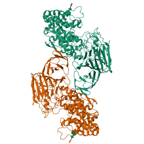
2FUQ

Crystal Structure of Heparinase II


Experimental Data Snapshot

  • Method: X-RAY DIFFRACTION
  • Resolution: 2.15 Å
  • R-Value Free: 0.235 
  • R-Value Work: 0.195 
  • R-Value Observed: 0.197 

wwPDB Validation   3D Report Full Report


This is version 3.2 of the entry. See complete history


Literature

Crystal Structure of Heparinase II from Pedobacter heparinus and Its Complex with a Disaccharide Product.

Shaya, D.Tocilj, A.Li, Y.Myette, J.Venkataraman, G.Sasisekharan, R.Cygler, M.

(2006) J Biol Chem 281: 15525-15535

  • DOI: https://doi.org/10.1074/jbc.M512055200
  • Primary Citation of Related Structures:  
    2FUQ, 2FUT

  • PubMed Abstract: 

    Heparinase II depolymerizes heparin and heparan sulfate glycosaminoglycans, yielding unsaturated oligosaccharide products through an elimination degradation mechanism. This enzyme cleaves the oligosaccharide chain on the nonreducing end of either glucuronic or iduronic acid, sharing this characteristic with a chondroitin ABC lyase. We have determined the first structure of a heparin-degrading lyase, that of heparinase II from Pedobacter heparinus (formerly Flavobacterium heparinum), in a ligand-free state at 2.15 A resolution and in complex with a disaccharide product of heparin degradation at 2.30 A resolution. The protein is composed of three domains: an N-terminal alpha-helical domain, a central two-layered beta-sheet domain, and a C-terminal domain forming a two-layered beta-sheet. Heparinase II shows overall structural similarities to the polysaccharide lyase family 8 (PL8) enzymes chondroitin AC lyase and hyaluronate lyase. In contrast to PL8 enzymes, however, heparinase II forms stable dimers, with the two active sites formed independently within each monomer. The structure of the N-terminal domain of heparinase II is also similar to that of alginate lyases from the PL5 family. A Zn2+ ion is bound within the central domain and plays an essential structural role in the stabilization of a loop forming one wall of the substrate-binding site. The disaccharide binds in a long, deep canyon formed at the top of the N-terminal domain and by loops extending from the central domain. Based on structural comparison with the lyases from the PL5 and PL8 families having bound substrates or products, the disaccharide found in heparinase II occupies the "+1" and "+2" subsites. The structure of the enzyme-product complex, combined with data from previously characterized mutations, allows us to propose a putative chemical mechanism of heparin and heparan-sulfate degradation.


  • Organizational Affiliation

    Department of Biochemistry, McGill University, Montreal, Quebec H3G 1Y6, Canada.


Macromolecules
Find similar proteins by:  (by identity cutoff)  |  3D Structure
Entity ID: 1
MoleculeChains Sequence LengthOrganismDetailsImage
heparinase II protein
A, B
747Pedobacter heparinusMutation(s): 2 
Gene Names: hep b
EC: 4.2.2.8 (UniProt), 4.2.2.7 (UniProt)
UniProt
Find proteins for C6XZB6 (Pedobacter heparinus (strain ATCC 13125 / DSM 2366 / CIP 104194 / JCM 7457 / NBRC 12017 / NCIMB 9290 / NRRL B-14731 / HIM 762-3))
Explore C6XZB6 
Go to UniProtKB:  C6XZB6
Entity Groups  
Sequence Clusters30% Identity50% Identity70% Identity90% Identity95% Identity100% Identity
UniProt GroupC6XZB6
Glycosylation
Glycosylation Sites: 1
Sequence Annotations
Expand
  • Reference Sequence
Oligosaccharides

Help

Entity ID: 2
MoleculeChains Length2D Diagram Glycosylation3D Interactions
alpha-D-xylopyranose-(1-4)-alpha-D-glucopyranuronic acid-(1-2)-[alpha-L-rhamnopyranose-(1-4)]alpha-D-mannopyranose
C, D
4O-Glycosylation
Glycosylation Resources
GlyTouCan:  G51375MF
GlyCosmos:  G51375MF
Small Molecules
Ligands 3 Unique
IDChains Name / Formula / InChI Key2D Diagram3D Interactions
PO4
Query on PO4

Download Ideal Coordinates CCD File 
F [auth A],
J [auth B]
PHOSPHATE ION
O4 P
NBIIXXVUZAFLBC-UHFFFAOYSA-K
ZN
Query on ZN

Download Ideal Coordinates CCD File 
E [auth A],
I [auth B]
ZINC ION
Zn
PTFCDOFLOPIGGS-UHFFFAOYSA-N
FMT
Query on FMT

Download Ideal Coordinates CCD File 
G [auth A],
H [auth A],
K [auth B]
FORMIC ACID
C H2 O2
BDAGIHXWWSANSR-UHFFFAOYSA-N
Modified Residues  1 Unique
IDChains TypeFormula2D DiagramParent
PCA
Query on PCA
A, B
L-PEPTIDE LINKINGC5 H7 N O3GLN
Experimental Data & Validation

Experimental Data

  • Method: X-RAY DIFFRACTION
  • Resolution: 2.15 Å
  • R-Value Free: 0.235 
  • R-Value Work: 0.195 
  • R-Value Observed: 0.197 
  • Space Group: P 21 21 21
Unit Cell:
Length ( Å )Angle ( ˚ )
a = 70.046α = 90
b = 119.339β = 90
c = 200.7γ = 90
Software Package:
Software NamePurpose
REFMACrefinement
HKL-2000data reduction
SCALEPACKdata scaling
SOLVEphasing

Structure Validation

View Full Validation Report


Entry History 

Deposition Data

Revision History  (Full details and data files)

  • Version 1.0: 2006-04-18
    Type: Initial release
  • Version 1.1: 2008-05-01
    Changes: Version format compliance
  • Version 1.2: 2011-07-13
    Changes: Advisory, Version format compliance
  • Version 1.3: 2016-03-23
    Changes: Derived calculations, Non-polymer description
  • Version 2.0: 2019-12-25
    Changes: Advisory, Data collection, Database references, Derived calculations, Polymer sequence
  • Version 3.0: 2020-07-29
    Type: Remediation
    Reason: Carbohydrate remediation
    Changes: Advisory, Atomic model, Data collection, Derived calculations, Structure summary
  • Version 3.1: 2021-10-20
    Changes: Database references, Derived calculations, Structure summary
  • Version 3.2: 2024-10-30
    Changes: Data collection, Structure summary