2FTK | pdb_00002ftk

berylloflouride Spo0F complex with Spo0B


Experimental Data Snapshot

  • Method: X-RAY DIFFRACTION
  • Resolution: 3.05 Å
  • R-Value Free: 
    0.282 (Depositor), 0.276 (DCC) 
  • R-Value Work: 
    0.232 (Depositor), 0.224 (DCC) 

Starting Model: experimental
View more details

wwPDB Validation 3D Report Full Report

Validation slider image for 2FTK

This is version 1.5 of the entry. See complete history

Literature

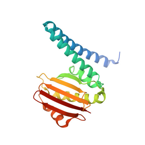
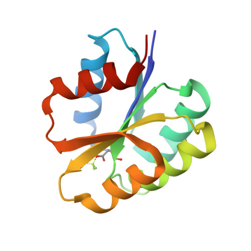
The Crystal Structure of Beryllofluoride Spo0F in Complex with the Phosphotransferase Spo0B Represents a Phosphotransfer Pretransition State.

Varughese, K.I.Tsigelny, I.Zhao, H.

(2006) J Bacteriol 188: 4970-4977

  • DOI: https://doi.org/10.1128/JB.00160-06
  • Primary Citation Related Structures: 
    2FTK

  • PubMed Abstract: 

    A number of regulatory circuits in biological systems function through the exchange of phosphoryl groups from one protein to another. Spo0F and Spo0B are components of a phosphorelay that control sporulation in the bacterium Bacillus subtilis through the exchange of a phosphoryl group. Using beryllofluoride as a mimic for phosphorylation, we trapped the interaction of the phosphorylated Spo0F with Spo0B in the crystal lattice. The transition state of phosphoryl transfer continues to be a highly debated issue, as to whether it is associative or dissociative in nature. The geometry of Spo0F binding to Spo0B favors an associative mechanism for phosphoryl transfer. In order to visualize the autophosphorylation of the histidine kinase, KinA, and the subsequent phosphoryl transfer to Spo0F, we generated in silico models representing these reaction steps.


  • Organizational Affiliation
    • Division of Cellular Biology, Department of Molecular and Experimental Medicine, MEM-116, The Scripps Research Institute, 10550 North Torrey Pines Road, La Jolla, CA 92037, USA. kiv@scripps.edu

Macromolecule Content 

  • Total Structure Weight: 147.33 kDa 
  • Atom Count: 9,834 
  • Modeled Residue Count: 1,202 
  • Deposited Residue Count: 1,264 
  • Unique protein chains: 2

Macromolecules

Find similar proteins by:|  3D Structure
Entity ID: 1
MoleculeChains  Sequence LengthOrganismDetailsImage
Sporulation initiation phosphotransferase B
A, B, C, D
192Bacillus subtilisMutation(s): 0 
Gene Names: spo0Bspo0D
EC: 2.7
UniProt
Find proteins for P06535 (Bacillus subtilis (strain 168))
Explore P06535 
Go to UniProtKB:  P06535
Entity Groups
Sequence Clusters30% Identity50% Identity70% Identity90% Identity95% Identity100% Identity
UniProt GroupP06535
Sequence Annotations
Expand
Reference Sequence
Find similar proteins by:|  3D Structure
Entity ID: 2
MoleculeChains  Sequence LengthOrganismDetailsImage
Sporulation initiation phosphotransferase F
E, F, G, H
124Bacillus subtilisMutation(s): 2 
Gene Names: spo0F
EC: 2.7
UniProt
Find proteins for P06628 (Bacillus subtilis (strain 168))
Explore P06628 
Go to UniProtKB:  P06628
Entity Groups
Sequence Clusters30% Identity50% Identity70% Identity90% Identity95% Identity100% Identity
UniProt GroupP06628
Sequence Annotations
Expand
Reference Sequence

Experimental Data & Validation

Experimental Data

  • Method: X-RAY DIFFRACTION
  • Resolution: 3.05 Å
  • R-Value Free:  0.282 (Depositor), 0.276 (DCC) 
  • R-Value Work:  0.232 (Depositor), 0.224 (DCC) 
Space Group: P 21 21 21
Unit Cell:
Length ( Å )Angle ( ˚ )
a = 73.467α = 90
b = 118.331β = 90
c = 168.235γ = 90
Software Package:
Software NamePurpose
Blu-Icedata collection
XDSdata reduction
CNSrefinement
XDSdata scaling
CNSphasing

Structure Validation

View Full Validation Report


Entry History 

Deposition Data

Revision History  (Full details and data files)

  • Version 1.0: 2006-07-18
    Type: Initial release
  • Version 1.1: 2008-05-01
    Changes: Version format compliance
  • Version 1.2: 2011-07-13
    Changes: Version format compliance
  • Version 1.3: 2021-10-20
    Changes: Database references, Derived calculations
  • Version 1.4: 2023-08-30
    Changes: Data collection, Refinement description
  • Version 1.5: 2024-11-20
    Changes: Structure summary