2FR6 | pdb_00002fr6

Crystal Structure of Mouse Cytidine Deaminase Complexed with Cytidine


Experimental Data Snapshot

  • Method: X-RAY DIFFRACTION
  • Resolution: 2.07 Å
  • R-Value Free: 
    0.214 (Depositor), 0.203 (DCC) 
  • R-Value Work: 
    0.188 (Depositor), 0.181 (DCC) 

Starting Model: experimental
View more details

wwPDB Validation 3D Report Full Report

Validation slider image for 2FR6

Ligand Structure Quality Assessment 


This is version 1.4 of the entry. See complete history

Literature

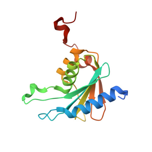
The 1.48 A Resolution Crystal Structure of the Homotetrameric Cytidine Deaminase from Mouse

Teh, A.H.Kimura, M.Yamamoto, M.Tanaka, N.Yamaguchi, I.Kumasaka, T.

(2006) Biochemistry 45: 7825-7833

  • DOI: https://doi.org/10.1021/bi060345f
  • Primary Citation Related Structures: 
    1ZAB, 2FR5, 2FR6

  • PubMed Abstract: 

    Cytidine deaminase (CDA) is a zinc-dependent enzyme that catalyzes the deamination of cytidine or deoxycytidine to form uridine or deoxyuridine. Here we present the crystal structure of mouse CDA (MmCDA), complexed with either tetrahydrouridine (THU), 3-deazauridine (DAU), or cytidine. In the MmCDA-DAU complex, it clearly demonstrates that cytidine is distinguished from uridine by its 4-NH(2) group that acts as a hydrogen bond donor. In the MmCDA-cytidine complex, cytidine, unexpectedly, binds as the substrate instead of the deaminated product in three of the four subunits, and in the remaining subunit it binds as the product uridine. Furthermore, the charge-neutralizing Arg68 of MmCDA has also exhibited two alternate conformations, I and II. In conformation I, the only conformation observed in the other structurally known homotetrameric CDAs, Arg68 hydrogen bonds Cys65 and Cys102 to modulate part of their negative charges. However, in conformation II the side chain of Arg68 rotates about 130 degrees around the Cgamma-Cdelta bond and abolishes these hydrogen bonds. The lack of hydrogen bonding may indirectly weaken the zinc-product interaction by increased electron donation from cysteine to the zinc ion, suggesting a novel product-expelling mechanism. On the basis of known structures, structural analysis further reveals two subclasses of homotetrameric CDAs that can be identified according to the position of the charge-neutralizing arginine residue. Implications for CDA-RNA interaction have also been considered.


  • Organizational Affiliation
    • Department of Life Science, Tokyo Institute of Technology, 4259-B-6 Nagatsuta-cho, Midori-ku, Yokohama, Kanagawa 226-8501, Japan.

Macromolecule Content 

  • Total Structure Weight: 65.94 kDa 
  • Atom Count: 4,691 
  • Modeled Residue Count: 543 
  • Deposited Residue Count: 584 
  • Unique protein chains: 1

Macromolecules

Find similar proteins by:|  3D Structure
Entity ID: 1
MoleculeChains  Sequence LengthOrganismDetailsImage
Cytidine deaminase
A, B, C, D
146Mus musculusMutation(s): 0 
EC: 3.5.4.5
UniProt
Find proteins for P56389 (Mus musculus)
Explore P56389 
Go to UniProtKB:  P56389
Entity Groups
Sequence Clusters30% Identity50% Identity70% Identity90% Identity95% Identity100% Identity
UniProt GroupP56389
Sequence Annotations
Expand
Reference Sequence

Small Molecules

Ligands 5 Unique
IDChains Name / Formula / InChI Key2D Diagram3D Interactions
URI

Query on URI


Download:Ideal Coordinates CCD File
I [auth B]URIDINE
C9 H12 N2 O6
DRTQHJPVMGBUCF-XVFCMESISA-N
CTN

Query on CTN


Download:Ideal Coordinates CCD File
G [auth A],
L [auth C],
N [auth D]
4-AMINO-1-BETA-D-RIBOFURANOSYL-2(1H)-PYRIMIDINONE
C9 H13 N3 O5
UHDGCWIWMRVCDJ-XVFCMESISA-N
SO4

Query on SO4


Download:Ideal Coordinates CCD File
F [auth A]SULFATE ION
O4 S
QAOWNCQODCNURD-UHFFFAOYSA-L
ZN

Query on ZN


Download:Ideal Coordinates CCD File
E [auth A],
H [auth B],
K [auth C],
M [auth D]
ZINC ION
Zn
PTFCDOFLOPIGGS-UHFFFAOYSA-N
NH3

Query on NH3


Download:Ideal Coordinates CCD File
J [auth B]AMMONIA
H3 N
QGZKDVFQNNGYKY-UHFFFAOYSA-N

Experimental Data & Validation

Experimental Data

  • Method: X-RAY DIFFRACTION
  • Resolution: 2.07 Å
  • R-Value Free:  0.214 (Depositor), 0.203 (DCC) 
  • R-Value Work:  0.188 (Depositor), 0.181 (DCC) 
Space Group: I 2 2 2
Unit Cell:
Length ( Å )Angle ( ˚ )
a = 82.199α = 90
b = 92.961β = 90
c = 180.737γ = 90
Software Package:
Software NamePurpose
d*TREKdata scaling
CNSrefinement
PDB_EXTRACTdata extraction

Structure Validation

View Full Validation Report


Ligand Structure Quality Assessment 


Entry History 

Deposition Data

  • Released Date: 2006-07-11 
  • Deposition Author(s): Teh, A.H.

Revision History  (Full details and data files)

  • Version 1.0: 2006-07-11
    Type: Initial release
  • Version 1.1: 2008-05-01
    Changes: Version format compliance
  • Version 1.2: 2011-07-13
    Changes: Version format compliance
  • Version 1.3: 2017-10-18
    Changes: Advisory, Refinement description
  • Version 1.4: 2023-10-25
    Changes: Advisory, Data collection, Database references, Derived calculations, Refinement description