2FQL | pdb_00002fql

Crystal structure of trimeric frataxin from the yeast Saccharomyces cerevisiae


Experimental Data Snapshot

  • Method: X-RAY DIFFRACTION
  • Resolution: 3.01 Å
  • R-Value Free: 
    0.305 (Depositor), 0.255 (DCC) 
  • R-Value Work: 
    0.286 (Depositor), 0.238 (DCC) 
  • R-Value Observed: 
    0.286 (Depositor) 

Starting Models: experimental
View more details

wwPDB Validation 3D Report Full Report

Validation slider image for 2FQL

This is version 1.5 of the entry. See complete history

Literature

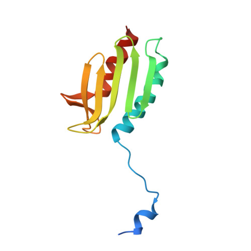
The structures of frataxin oligomers reveal the mechanism for the delivery and detoxification of iron.

Karlberg, T.Schagerlof, U.Gakh, O.Park, S.Ryde, U.Lindahl, M.Leath, K.Garman, E.Isaya, G.Al-Karadaghi, S.

(2006) Structure 14: 1535-1546

  • DOI: https://doi.org/10.1016/j.str.2006.08.010
  • Primary Citation Related Structures: 
    2FQL

  • PubMed Abstract: 

    Defects in the mitochondrial protein frataxin are responsible for Friedreich ataxia, a neurodegenerative and cardiac disease that affects 1:40,000 children. Here, we present the crystal structures of the iron-free and iron-loaded frataxin trimers, and a single-particle electron microscopy reconstruction of a 24 subunit oligomer. The structures reveal fundamental aspects of the frataxin mechanism. The trimer has a central channel in which one atom of iron binds. Two conformations of the channel with different metal-binding affinities suggest that a gating mechanism controls whether the bound iron is delivered to other proteins or transferred to detoxification sites. The trimer constitutes the basic structural unit of the 24 subunit oligomer. The architecture of this oligomer and several features of the trimer structure demonstrate striking similarities to the iron-storage protein ferritin. The data reveal how stepwise assembly provides frataxin with the structural flexibility to perform two functions: metal delivery and detoxification.


  • Organizational Affiliation
    • Department of Molecular Biophysics, Center for Chemistry and Chemical Engineering, Lund University, P.O. Box 124, SE-221 00 Lund, Sweden.

Macromolecule Content 

  • Total Structure Weight: 13.67 kDa 
  • Atom Count: 883 
  • Modeled Residue Count: 112 
  • Deposited Residue Count: 123 
  • Unique protein chains: 1

Macromolecules

Find similar proteins by:|  3D Structure
Entity ID: 1
MoleculeChains  Sequence LengthOrganismDetailsImage
Frataxin homolog, mitochondrial123Saccharomyces cerevisiaeMutation(s): 1 
Gene Names: YFH1
EC: 1.16.3.1
UniProt
Find proteins for Q07540 (Saccharomyces cerevisiae (strain ATCC 204508 / S288c))
Explore Q07540 
Go to UniProtKB:  Q07540
Entity Groups
Sequence Clusters30% Identity50% Identity70% Identity90% Identity95% Identity100% Identity
UniProt GroupQ07540
Sequence Annotations
Expand
Reference Sequence

Experimental Data & Validation

Experimental Data

  • Method: X-RAY DIFFRACTION
  • Resolution: 3.01 Å
  • R-Value Free:  0.305 (Depositor), 0.255 (DCC) 
  • R-Value Work:  0.286 (Depositor), 0.238 (DCC) 
  • R-Value Observed: 0.286 (Depositor) 
Space Group: I 21 3
Unit Cell:
Length ( Å )Angle ( ˚ )
a = 121.22α = 90
b = 121.22β = 90
c = 121.22γ = 90
Software Package:
Software NamePurpose
CNSrefinement
PDB_EXTRACTdata extraction
MAR345data collection
XDSdata scaling
PHASERphasing

Structure Validation

View Full Validation Report


Entry History 

Deposition Data

Revision History  (Full details and data files)

  • Version 1.0: 2006-11-07
    Type: Initial release
  • Version 1.1: 2008-05-01
    Changes: Version format compliance
  • Version 1.2: 2011-07-13
    Changes: Version format compliance
  • Version 1.3: 2017-10-18
    Changes: Advisory, Data collection, Refinement description
  • Version 1.4: 2021-10-20
    Changes: Advisory, Database references
  • Version 1.5: 2023-08-30
    Changes: Data collection, Refinement description