2FOP

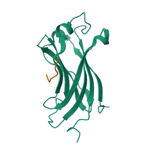
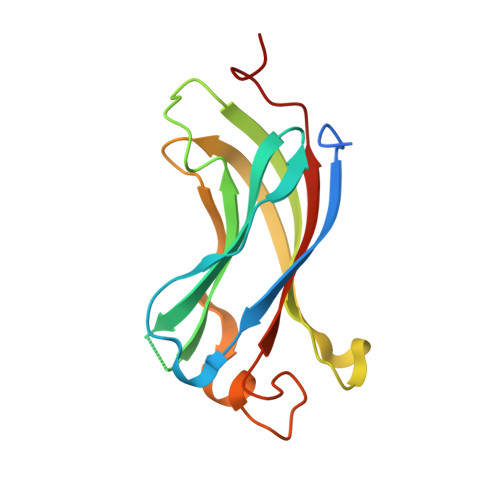
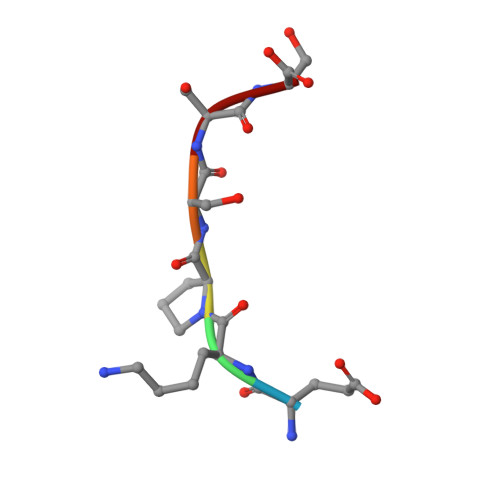
The Crystal Structure of the N-terminal domain of HAUSP/USP7 complexed with mdm2 peptide 147-150


Experimental Data Snapshot

  • Method: X-RAY DIFFRACTION
  • Resolution: 2.10 Å
  • R-Value Free: 0.250 
  • R-Value Work: 0.206 
  • R-Value Observed: 0.206 

Starting Model: experimental
View more details

wwPDB Validation   3D Report Full Report


This is version 1.4 of the entry. See complete history


Literature

Molecular recognition of p53 and MDM2 by USP7/HAUSP

Sheng, Y.Saridakis, V.Sarkari, F.Duan, S.Wu, T.Arrowsmith, C.H.Frappier, L.

(2006) Nat Struct Mol Biol 13: 285-291

  • DOI: https://doi.org/10.1038/nsmb1067
  • Primary Citation of Related Structures:  
    2FOJ, 2FOO, 2FOP

  • PubMed Abstract: 

    The ubiquitin-specific protease, USP7, has key roles in the p53 pathway whereby it stabilizes both p53 and MDM2. We show that the N-terminal domain of USP7 binds two closely spaced 4-residue sites in both p53 and MDM2, falling between p53 residues 359-367 and MDM2 residues 147-159. Cocrystal structures with USP7 were determined for both p53 peptides and for one MDM2 peptide. These peptides bind the same surface of USP7 as Epstein-Barr nuclear antigen-1, explaining the competitive nature of the interactions. The structures and mutagenesis data indicate a preference for a P/AXXS motif in peptides that bind USP7. Contacts made by serine are identical and crucial for all peptides, and Trp165 in the peptide-binding pocket of USP7 is also crucial. These results help to elucidate the mechanism of substrate recognition by USP7 and the regulation of the p53 pathway.


  • Organizational Affiliation

    Ontario Cancer Institute, Department of Medical Biophysics, 101 College Street, Toronto, Ontario, Canada M5G 1L7.


Macromolecules
Find similar proteins by:  (by identity cutoff)  |  3D Structure
Entity ID: 1
MoleculeChains Sequence LengthOrganismDetailsImage
Ubiquitin carboxyl-terminal hydrolase 7155Homo sapiensMutation(s): 0 
Gene Names: HAUSPUSP7
EC: 3.1.2.15 (PDB Primary Data), 3.4.19.12 (UniProt)
UniProt & NIH Common Fund Data Resources
Find proteins for Q93009 (Homo sapiens)
Explore Q93009 
Go to UniProtKB:  Q93009
PHAROS:  Q93009
GTEx:  ENSG00000187555 
Entity Groups  
Sequence Clusters30% Identity50% Identity70% Identity90% Identity95% Identity100% Identity
UniProt GroupQ93009
Sequence Annotations
Expand
  • Reference Sequence

Find similar proteins by:  Sequence   |   3D Structure  

Entity ID: 2
MoleculeChains Sequence LengthOrganismDetailsImage
mdm2 peptide6Homo sapiensMutation(s): 0 
Gene Names: MDM2
EC: 2.3.2.27
UniProt & NIH Common Fund Data Resources
Find proteins for Q00987 (Homo sapiens)
Explore Q00987 
Go to UniProtKB:  Q00987
PHAROS:  Q00987
GTEx:  ENSG00000135679 
Entity Groups  
UniProt GroupQ00987
Sequence Annotations
Expand
  • Reference Sequence
Experimental Data & Validation

Experimental Data

  • Method: X-RAY DIFFRACTION
  • Resolution: 2.10 Å
  • R-Value Free: 0.250 
  • R-Value Work: 0.206 
  • R-Value Observed: 0.206 
  • Space Group: P 41
Unit Cell:
Length ( Å )Angle ( ˚ )
a = 69.9α = 90
b = 69.9β = 90
c = 45.7γ = 90
Software Package:
Software NamePurpose
CNSrefinement
HKL-2000data reduction
SCALEPACKdata scaling
CNSphasing

Structure Validation

View Full Validation Report


Entry History 

Deposition Data

Revision History  (Full details and data files)

  • Version 1.0: 2006-02-14
    Type: Initial release
  • Version 1.1: 2008-05-01
    Changes: Version format compliance
  • Version 1.2: 2011-07-13
    Changes: Version format compliance
  • Version 1.3: 2016-12-21
    Changes: Structure summary
  • Version 1.4: 2023-08-30
    Changes: Data collection, Database references, Refinement description