2FN2 | pdb_00002fn2

SOLUTION NMR STRUCTURE OF THE GLYCOSYLATED SECOND TYPE TWO MODULE OF FIBRONECTIN, 20 STRUCTURES


Experimental Data Snapshot

  • Method: SOLUTION NMR
  • Conformers Calculated: 80 
  • Conformers Submitted: 20 
  • Selection Criteria: ENERGY, AGREEMENT WITH EXPERIMENTAL DATA 

wwPDB Validation 3D Report Full Report

Validation slider image for 2FN2

This is version 1.4 of the entry. See complete history

Literature

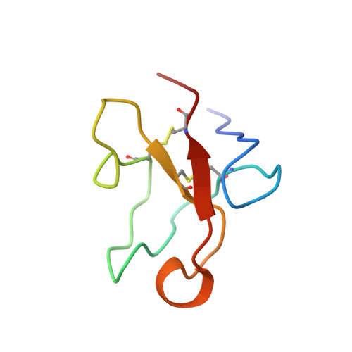
Solution structure of the glycosylated second type 2 module of fibronectin.

Sticht, H.Pickford, A.R.Potts, J.R.Campbell, I.D.

(1998) J Mol Biology 276: 177-187

  • DOI: https://doi.org/10.1006/jmbi.1997.1528
  • Primary Citation Related Structures: 
    2FN2

  • PubMed Abstract: 

    Fibronectin is an extracellular matrix glycoprotein that plays a role in a number of physiological processes involving cell adhesion and migration. The modules of the fibronectin monomer are organized into proteolytically resistant domains that in isolation retain their affinity for various ligands. The tertiary structure of the glycosylated second type 2 module (2F2) from the gelatin-binding domain of fibronectin was determined by two-dimensional nuclear magnetic resonance spectroscopy and simulated annealing. The structure is well defined with an overall fold typical of F2 modules, showing two double-stranded antiparallel beta-sheets and a partially solvent-exposed hydrophobic cluster. An N-terminal beta-sheet, that was not present in previously determined F2 module structures, may be important for defining the relative orientation of adjacent F2 modules in fibronectin. This is the first three-dimensional structure of a glycosylated module of fibronectin, and provides insight into the possible role of the glycosylation in protein stability, protease resistance and modulation of collagen binding. Based on the structures of the isolated modules, models for the 1F22F2 pair were generated by randomly changing the orientation of the linker peptide between the modules. The models suggest that the two putative collagen binding sites in the pair form discrete binding sites, rather than combining to form a single binding site.


  • Organizational Affiliation
    • Department of Biochemistry University of Oxford, UK.

Macromolecule Content 

  • Total Structure Weight: 6.92 kDa 
  • Atom Count: 482 
  • Modeled Residue Count: 59 
  • Deposited Residue Count: 59 
  • Unique protein chains: 1

Macromolecules

Find similar proteins by:|  3D Structure
Entity ID: 1
MoleculeChains  Sequence LengthOrganismDetailsImage
FIBRONECTIN59Homo sapiensMutation(s): 0 
UniProt & NIH Common Fund Data Resources
Find proteins for P02751 (Homo sapiens)
Explore P02751 
Go to UniProtKB:  P02751
PHAROS:  P02751
GTEx:  ENSG00000115414 
Entity Groups
Sequence Clusters30% Identity50% Identity70% Identity90% Identity95% Identity100% Identity
UniProt GroupP02751
Glycosylation
Glycosylation Sites: 1Go to GlyGen: P02751-15
Sequence Annotations
Expand
Reference Sequence

Small Molecules

Ligands 1 Unique
IDChains Name / Formula / InChI Key2D Diagram3D Interactions
NAG

Query on NAG


Download:Ideal Coordinates CCD File
B [auth A]2-acetamido-2-deoxy-beta-D-glucopyranose
C8 H15 N O6
OVRNDRQMDRJTHS-FMDGEEDCSA-N

Experimental Data & Validation

Experimental Data

  • Method: SOLUTION NMR
  • Conformers Calculated: 80 
  • Conformers Submitted: 20 
  • Selection Criteria: ENERGY, AGREEMENT WITH EXPERIMENTAL DATA 

Structure Validation

View Full Validation Report


Entry History 

Deposition Data

Revision History  (Full details and data files)

  • Version 1.0: 1998-09-16
    Type: Initial release
  • Version 1.1: 2008-03-24
    Changes: Version format compliance
  • Version 1.2: 2011-07-13
    Changes: Non-polymer description, Version format compliance
  • Version 1.3: 2020-07-29
    Type: Remediation
    Reason: Carbohydrate remediation
    Changes: Data collection, Derived calculations, Other, Structure summary
  • Version 1.4: 2024-10-30
    Changes: Data collection, Database references, Structure summary