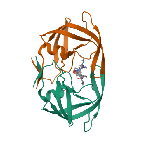
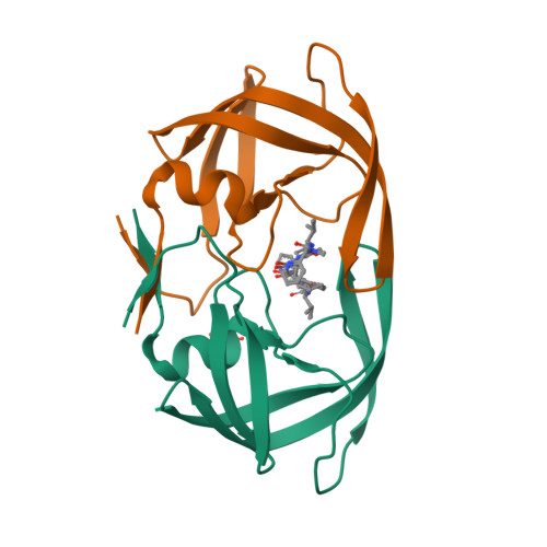
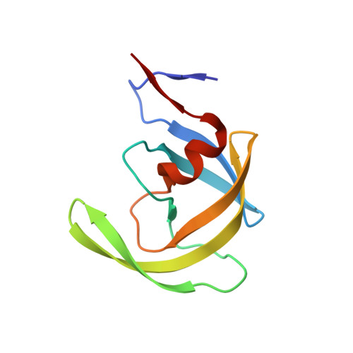
2FLE | pdb_00002fle

Structural analysis of asymmetric inhibitor bound to the HIV-1 Protease V82A mutant


Experimental Data Snapshot

  • Method: X-RAY DIFFRACTION
  • Resolution: 1.90 Å
  • R-Value Free: 
    0.251 (Depositor) 
  • R-Value Work: 
    0.199 (Depositor), 0.200 (DCC) 
  • R-Value Observed: 
    0.207 (Depositor) 

Starting Model: experimental
View more details

wwPDB Validation   3D Report Full Report


Ligand Structure Quality Assessment 

Created with Raphaël 2.3.0Worse 01 BetterLigand structure goodness of fit to experimental dataBest fitted AIClick on this verticalbar to view details

This is version 1.5 of the entry. See complete history


Literature

Design, synthesis, evaluation, and crystallographic-based structural studies of HIV-1 protease inhibitors with reduced response to the V82A mutation.

Clemente, J.C.Robbins, A.Grana, P.Paleo, M.R.Correa, J.F.Villaverde, M.C.Sardina, F.J.Govindasamy, L.Agbandje-McKenna, M.McKenna, R.Dunn, B.M.Sussman, F.

(2008) J Med Chem 51: 852-860

  • DOI: https://doi.org/10.1021/jm701170f
  • Primary Citation of Related Structures:  
    2FLE

  • PubMed Abstract: 

    In our quest for HIV-1 protease inhibitors that are not affected by the V82A resistance mutation, we have synthesized and tested a second generation set of C2-symmetric HIV-1 protease inhibitors that contain a cyclohexane group at P1 and/or P1'. The binding affinity results indicate that these compounds have an improved response to the appearance of the V82A mutation than the parent compound. The X-ray structure of one of these compounds with the V82A HIV-1 PR variant provides the structural rationale for the better resistance profile of these compounds. Moreover, scrutiny of the X-ray structure suggests that the ring of the Cha side chain might be in a boat rather than in the chair conformation, a result supported by molecular dynamics simulations.


  • Organizational Affiliation

    Department of Biochemistry and Molecular Biology, University of Florida College of Medicine, Gainesville, Florida 32610, USA.


Macromolecules
Find similar proteins by:  (by identity cutoff)  |  3D Structure
Entity ID: 1
MoleculeChains Sequence LengthOrganismDetailsImage
pol protein
A, B
99Human immunodeficiency virus 1Mutation(s): 1 
UniProt
Find proteins for Q9J2R0 (Human immunodeficiency virus type 1)
Explore Q9J2R0 
Go to UniProtKB:  Q9J2R0
Entity Groups  
Sequence Clusters30% Identity50% Identity70% Identity90% Identity95% Identity100% Identity
UniProt GroupQ9J2R0
Sequence Annotations
Expand
  • Reference Sequence
Small Molecules
Ligands 2 Unique
IDChains Name / Formula / InChI Key2D Diagram3D Interactions
AI
Query on AI

Download Ideal Coordinates CCD File 
D [auth B](2S,2'S)-N,N'-[(2S,3S,4S,5S)-1-CYCLOHEXYL-3,4-DIHYDROXY-6-PHENYLHEXANE-2,5-DIYL]BIS[3-METHYL-2-({[METHYL(PYRIDIN-2-YLMETHYL)AMINO]CARBONYL}AMINO)BUTANAMIDE]
C44 H64 N8 O6
JQIFSYRTTKZQMY-UNHORJANSA-N
GOL
Query on GOL

Download Ideal Coordinates CCD File 
C [auth A]GLYCEROL
C3 H8 O3
PEDCQBHIVMGVHV-UHFFFAOYSA-N
Binding Affinity Annotations 
IDSourceBinding Affinity
AI BindingDB:  2FLE Ki: min: 3.2, max: 14 (nM) from 2 assay(s)
Experimental Data & Validation

Experimental Data

  • Method: X-RAY DIFFRACTION
  • Resolution: 1.90 Å
  • R-Value Free:  0.251 (Depositor) 
  • R-Value Work:  0.199 (Depositor), 0.200 (DCC) 
  • R-Value Observed: 0.207 (Depositor) 
Space Group: P 61
Unit Cell:
Length ( Å )Angle ( ˚ )
a = 62.195α = 90
b = 62.195β = 90
c = 82.752γ = 120
Software Package:
Software NamePurpose
DENZOdata reduction
SCALEPACKdata scaling
CNSrefinement
PDB_EXTRACTdata extraction
HKL-2000data reduction
CNSphasing

Structure Validation

View Full Validation Report


Ligand Structure Quality Assessment 

Created with Raphaël 2.3.0Worse 01 BetterLigand structure goodness of fit to experimental dataBest fitted AIClick on this verticalbar to view details

Entry History 

Deposition Data

Revision History  (Full details and data files)

  • Version 1.0: 2007-01-16
    Type: Initial release
  • Version 1.1: 2008-05-01
    Changes: Version format compliance
  • Version 1.2: 2011-07-13
    Changes: Non-polymer description, Version format compliance
  • Version 1.3: 2017-10-18
    Changes: Refinement description
  • Version 1.4: 2021-10-20
    Changes: Database references, Derived calculations
  • Version 1.5: 2023-08-30
    Changes: Data collection, Refinement description