2FFR | pdb_00002ffr

Crystallographic studies on N-azido-beta-D-glucopyranosylamine, an inhibitor of glycogen phosphorylase: comparison with N-acetyl-beta-D-glucopyranosylamine


Experimental Data Snapshot

  • Method: X-RAY DIFFRACTION
  • Resolution: 2.03 Å
  • R-Value Free: 
    0.199 (Depositor) 
  • R-Value Work: 
    0.184 (Depositor) 
  • R-Value Observed: 
    0.184 (Depositor) 

Starting Model: experimental
View more details

wwPDB Validation 3D Report Full Report

Validation slider image for 2FFR

This is version 1.4 of the entry. See complete history

Literature

Crystallographic studies on N-azidoacetyl-beta-d-glucopyranosylamine, an inhibitor of glycogen phosphorylase: Comparison with N-acetyl-beta-d-glucopyranosylamine.

Petsalakis, E.I.Chrysina, E.D.Tiraidis, C.Hadjiloi, T.Leonidas, D.D.Oikonomakos, N.G.Aich, U.Varghese, B.Loganathan, D.

(2006) Bioorg Med Chem 14: 5316-5324

  • DOI: https://doi.org/10.1016/j.bmc.2006.03.044
  • Primary Citation Related Structures: 
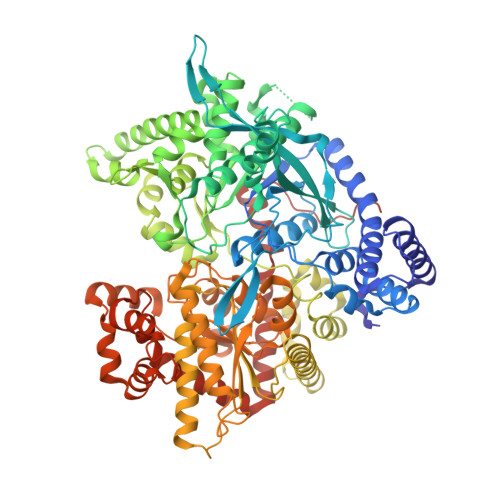
    2FFR

  • PubMed Abstract: 

    N-acetyl-beta-D-glucopyranosylamine (NAG) is a potent inhibitor (Ki=32 microM) of glycogen phosphorylase b (GPb), and has been employed as a lead compound for the structure-based design of new analogues, in an effort to utilize its potential as a hypoglycaemic agent. Replacement of the acetamido group by azidoacetamido group resulted in an inhibitor, N-azidoacetyl-beta-D-glucopyranosylamine (azido-NAG), with a Ki value of 48.7 microM, in the direction of glycogen synthesis. In order to elucidate the mechanism of inhibition, we determined the ligand structure in complex with GPb at 2.03 A resolution, and the structure of the fully acetylated derivative in the free form. The molecular packing of the latter is stabilized by a number of bifurcated hydrogen bonds of which the one involving a bifurcated C-H...N...H-C type hydrogen bonding is rather unique in organic azides. Azido-NAG can be accommodated in the catalytic site of T-state GPb at approximately the same position as that of NAG and stabilizes the T-state conformation of the 280 s loop by making several favourable contacts to residues of this loop. The difference observed in the Ki values of the two analogues can be interpreted in terms of desolvation effects, subtle structural changes of protein residues and changes in water structure.


  • Organizational Affiliation
    • Institute of Organic and Pharmaceutical Chemistry, The National Hellenic Research Foundation, 48, Vas. Constantinou Ave., 116 35 Athens, Greece.

Macromolecule Content 

  • Total Structure Weight: 95.79 kDa 
  • Atom Count: 6,981 
  • Modeled Residue Count: 810 
  • Deposited Residue Count: 825 
  • Unique protein chains: 1

Macromolecules

Find similar proteins by:|  3D Structure
Entity ID: 1
MoleculeChains  Sequence LengthOrganismDetailsImage
Glycogen phosphorylase, muscle form825Oryctolagus cuniculusMutation(s): 0 
EC: 2.4.1.1
UniProt
Find proteins for P00489 (Oryctolagus cuniculus)
Explore P00489 
Go to UniProtKB:  P00489
Entity Groups
Sequence Clusters30% Identity50% Identity70% Identity90% Identity95% Identity100% Identity
UniProt GroupP00489
Sequence Annotations
Expand
Reference Sequence

Small Molecules

Ligands 2 Unique
IDChains Name / Formula / InChI Key2D Diagram3D Interactions
DL6

Query on DL6


Download:Ideal Coordinates CCD File
C [auth A]N-(azidoacetyl)-beta-D-glucopyranosylamine
C8 H14 N4 O6
ACLUEOOMMLAETN-RHROMQPHSA-N
PLP

Query on PLP


Download:Ideal Coordinates CCD File
B [auth A]PYRIDOXAL-5'-PHOSPHATE
C8 H10 N O6 P
NGVDGCNFYWLIFO-UHFFFAOYSA-N

Experimental Data & Validation

Experimental Data

  • Method: X-RAY DIFFRACTION
  • Resolution: 2.03 Å
  • R-Value Free:  0.199 (Depositor) 
  • R-Value Work:  0.184 (Depositor) 
  • R-Value Observed: 0.184 (Depositor) 
Space Group: P 43 21 2
Unit Cell:
Length ( Å )Angle ( ˚ )
a = 128.626α = 90
b = 128.626β = 90
c = 116.163γ = 90
Software Package:
Software NamePurpose
CNSrefinement
HKL-2000data reduction
SCALEPACKdata scaling
CNSphasing

Structure Validation

View Full Validation Report


Entry History 

Deposition Data

Revision History  (Full details and data files)

  • Version 1.0: 2006-06-20
    Type: Initial release
  • Version 1.1: 2008-05-01
    Changes: Version format compliance
  • Version 1.2: 2011-07-13
    Changes: Derived calculations, Version format compliance
  • Version 1.3: 2020-07-29
    Type: Remediation
    Reason: Carbohydrate remediation
    Changes: Database references, Derived calculations, Structure summary
  • Version 1.4: 2023-08-30
    Changes: Data collection, Database references, Refinement description, Structure summary