2FEA | pdb_00002fea

Crystal structure of MtnX phosphatase from Bacillus Subtilis at 2.00 A resolution


Experimental Data Snapshot

  • Method: X-RAY DIFFRACTION
  • Resolution: 2.00 Å
  • R-Value Free: 
    0.217 (Depositor), 0.226 (DCC) 
  • R-Value Work: 
    0.160 (Depositor), 0.172 (DCC) 
  • R-Value Observed: 
    0.163 (Depositor) 

wwPDB Validation 3D Report Full Report

Validation slider image for 2FEA

This is version 1.4 of the entry. See complete history

Literature

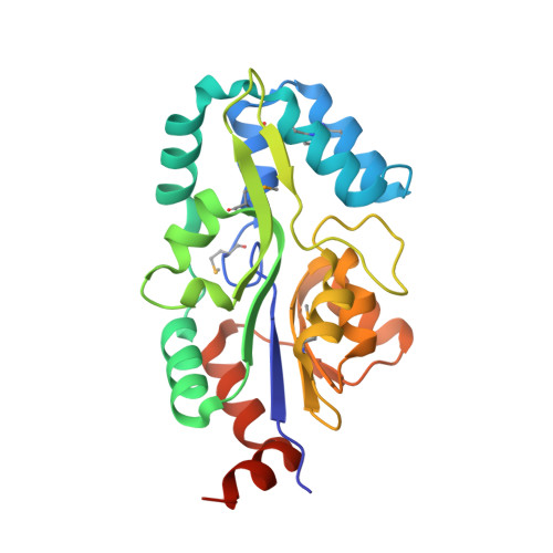
Crystal structure of MtnX phosphatase from Bacillus subtilis at 2.0 A resolution provides a structural basis for bipartite phosphomonoester hydrolysis of 2-hydroxy-3-keto-5-methylthiopentenyl-1-phosphate.

Xu, Q.Saikatendu, K.S.Krishna, S.S.McMullan, D.Abdubek, P.Agarwalla, S.Ambing, E.Astakhova, T.Axelrod, H.L.Carlton, D.Chiu, H.J.Clayton, T.DiDonato, M.Duan, L.Elsliger, M.A.Feuerhelm, J.Grzechnik, S.K.Hale, J.Hampton, E.Han, G.W.Haugen, J.Jaroszewski, L.Jin, K.K.Klock, H.E.Knuth, M.W.Koesema, E.Miller, M.D.Morse, A.T.Nigoghossian, E.Okach, L.Oommachen, S.Paulsen, J.Reyes, R.Rife, C.L.Schwarzenbacher, R.van den Bedem, H.White, A.Wolf, G.Hodgson, K.O.Wooley, J.Deacon, A.M.Godzik, A.Lesley, S.A.Wilson, I.A.

(2007) Proteins 69: 433-439

Macromolecule Content 

  • Total Structure Weight: 55.63 kDa 
  • Atom Count: 4,061 
  • Modeled Residue Count: 450 
  • Deposited Residue Count: 472 
  • Unique protein chains: 1

Macromolecules

Find similar proteins by:|  3D Structure
Entity ID: 1
MoleculeChains  Sequence LengthOrganismDetailsImage
2-hydroxy-3-keto-5-methylthiopentenyl-1-phosphate phosphatase
A, B
236Bacillus subtilisMutation(s): 0 
Gene Names: mtnX
EC: 3.1.3 (PDB Primary Data), 3.1.3.87 (UniProt)
UniProt
Find proteins for O31667 (Bacillus subtilis (strain 168))
Explore O31667 
Go to UniProtKB:  O31667
Entity Groups
Sequence Clusters30% Identity50% Identity70% Identity90% Identity95% Identity100% Identity
UniProt GroupO31667
Sequence Annotations
Expand
Reference Sequence

Small Molecules

Ligands 3 Unique
IDChains Name / Formula / InChI Key2D Diagram3D Interactions
ZN

Query on ZN


Download:Ideal Coordinates CCD File
D [auth A],
J [auth B]
ZINC ION
Zn
PTFCDOFLOPIGGS-UHFFFAOYSA-N
EDO

Query on EDO


Download:Ideal Coordinates CCD File
E [auth A]
F [auth A]
G [auth A]
H [auth A]
K [auth B]
E [auth A],
F [auth A],
G [auth A],
H [auth A],
K [auth B],
L [auth B],
M [auth B],
N [auth B],
O [auth B],
P [auth B]
1,2-ETHANEDIOL
C2 H6 O2
LYCAIKOWRPUZTN-UHFFFAOYSA-N
MG

Query on MG


Download:Ideal Coordinates CCD File
C [auth A],
I [auth B]
MAGNESIUM ION
Mg
JLVVSXFLKOJNIY-UHFFFAOYSA-N
Modified Residues  1 Unique
IDChains TypeFormula2D DiagramParent
MSE
Query on MSE
A, B
L-PEPTIDE LINKINGC5 H11 N O2 SeMET

Experimental Data & Validation

Experimental Data

  • Method: X-RAY DIFFRACTION
  • Resolution: 2.00 Å
  • R-Value Free:  0.217 (Depositor), 0.226 (DCC) 
  • R-Value Work:  0.160 (Depositor), 0.172 (DCC) 
  • R-Value Observed: 0.163 (Depositor) 
Space Group: P 1 21 1
Unit Cell:
Length ( Å )Angle ( ˚ )
a = 59.013α = 90
b = 43.104β = 98.91
c = 109.553γ = 90
Software Package:
Software NamePurpose
REFMACrefinement
SCALAdata scaling
PDB_EXTRACTdata extraction
MOSFLMdata reduction
CCP4data scaling
SOLVEphasing

Structure Validation

View Full Validation Report


Entry History 

Deposition Data

Revision History  (Full details and data files)

  • Version 1.0: 2005-12-27
    Type: Initial release
  • Version 1.1: 2008-05-01
    Changes: Version format compliance
  • Version 1.2: 2011-07-13
    Changes: Advisory, Version format compliance
  • Version 1.3: 2023-01-25
    Changes: Database references, Derived calculations
  • Version 1.4: 2024-11-06
    Changes: Data collection, Refinement description, Structure summary