2F3K | pdb_00002f3k

Substrate envelope and drug resistance: crystal structure of r01 in complex with wild-type hiv-1 protease


Experimental Data Snapshot

  • Method: X-RAY DIFFRACTION
  • Resolution: 1.60 Å
  • R-Value Free: 
    0.197 (Depositor), 0.206 (DCC) 
  • R-Value Work: 
    0.163 (Depositor), 0.175 (DCC) 
  • R-Value Observed: 
    0.165 (Depositor) 

Starting Model: experimental
View more details

wwPDB Validation 3D Report Full Report

Validation slider image for 2F3K

Ligand Structure Quality Assessment 


This is version 1.5 of the entry. See complete history

Literature

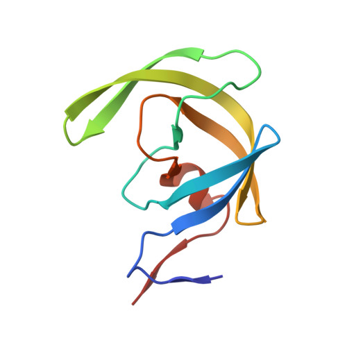
Substrate envelope and drug resistance: crystal structure of RO1 in complex with wild-type human immunodeficiency virus type 1 protease.

Prabu-Jeyabalan, M.King, N.M.Nalivaika, E.A.Heilek-Snyder, G.Cammack, N.Schiffer, C.A.

(2006) Antimicrob Agents Chemother 50: 1518-1521

  • DOI: https://doi.org/10.1128/AAC.50.4.1518-1521.2006
  • Primary Citation Related Structures: 
    2F3K

  • PubMed Abstract: 

    In our previous crystallographic studies of human immunodeficiency virus type 1 (HIV-1) protease-substrate complexes, we described a conserved "envelope" that appears to be important for substrate recognition and the selection of drug-resistant mutations. In this study, the complex of HIV-1 protease with the inhibitor RO1 was determined and comparison with the substrate envelope provides a rationale for mutational patterns.


  • Organizational Affiliation
    • Department of Biochemistry & Molecular Pharmacology, University of Massachusetts Medical School, 364 Plantation St., Worcester, MA 01605, USA.

Macromolecule Content 

  • Total Structure Weight: 22.82 kDa 
  • Atom Count: 2,168 
  • Modeled Residue Count: 198 
  • Deposited Residue Count: 198 
  • Unique protein chains: 1

Macromolecules

Find similar proteins by:|  3D Structure
Entity ID: 1
MoleculeChains  Sequence LengthOrganismDetailsImage
Protease
A, B
99Human immunodeficiency virus 1Mutation(s): 1 
EC: 3.4.23.16
UniProt
Find proteins for P03369 (Human immunodeficiency virus type 1 group M subtype B (isolate ARV2/SF2))
Explore P03369 
Go to UniProtKB:  P03369
Entity Groups
Sequence Clusters30% Identity50% Identity70% Identity90% Identity95% Identity100% Identity
UniProt GroupP03369
Sequence Annotations
Expand
Reference Sequence

Experimental Data & Validation

Experimental Data

  • Method: X-RAY DIFFRACTION
  • Resolution: 1.60 Å
  • R-Value Free:  0.197 (Depositor), 0.206 (DCC) 
  • R-Value Work:  0.163 (Depositor), 0.175 (DCC) 
  • R-Value Observed: 0.165 (Depositor) 
Space Group: P 21 21 21
Unit Cell:
Length ( Å )Angle ( ˚ )
a = 50.873α = 90
b = 58.167β = 90
c = 61.653γ = 90
Software Package:
Software NamePurpose
REFMACrefinement
SCALEPACKdata scaling
AMoREphasing

Structure Validation

View Full Validation Report


Ligand Structure Quality Assessment 


Entry History 

Deposition Data

Revision History  (Full details and data files)

  • Version 1.0: 2006-11-21
    Type: Initial release
  • Version 1.1: 2008-05-01
    Changes: Version format compliance
  • Version 1.2: 2011-07-13
    Changes: Version format compliance
  • Version 1.3: 2017-10-18
    Changes: Refinement description
  • Version 1.4: 2021-10-20
    Changes: Database references, Derived calculations
  • Version 1.5: 2023-08-23
    Changes: Data collection, Refinement description