2F32

Xray crystal structure of lysozyme mutant L20/R63A liganded to ethylguanidinium


Experimental Data Snapshot

  • Method: X-RAY DIFFRACTION
  • Resolution: 1.80 Å
  • R-Value Free: 0.245 
  • R-Value Work: 0.200 
  • R-Value Observed: 0.200 

Starting Model: experimental
View more details

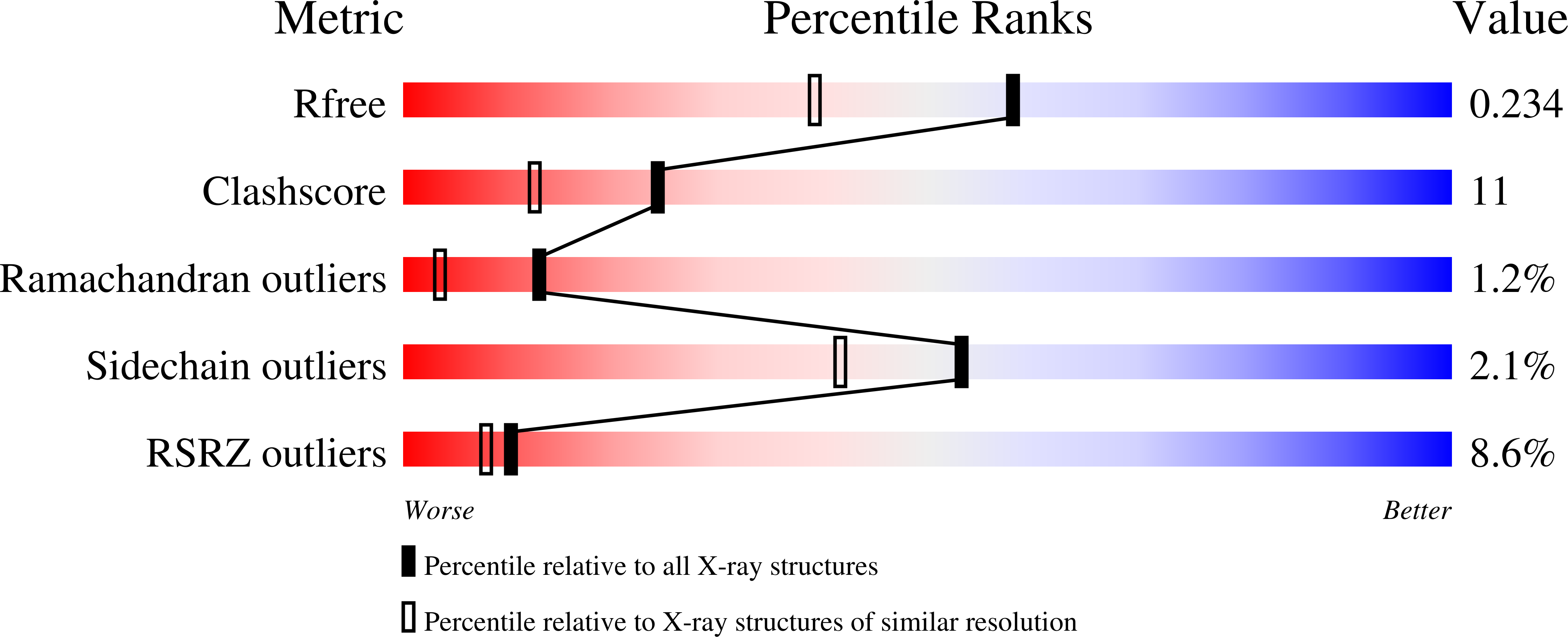
wwPDB Validation   3D Report Full Report


This is version 1.5 of the entry. See complete history


Literature

Guanidinium derivatives bind preferentially and trigger long-distance conformational changes in an engineered T4 lysozyme.

Yousef, M.S.Bischoff, N.Dyer, C.M.Baase, W.A.Matthews, B.W.

(2006) Protein Sci 15: 853-861

  • DOI: https://doi.org/10.1110/ps.052020606
  • Primary Citation of Related Structures:  
    2F2Q, 2F32, 2F47

  • PubMed Abstract: 

    The binding of guanidinium ion has been shown to promote a large-scale translation of a tandemly duplicated helix in an engineered mutant of T4 lysozyme. The guanidinium ion acts as a surrogate for the guanidino group of an arginine side chain. Here we determine whether methyl- and ethylguanidinium provide better mimics. The results show that addition of the hydrophobic moieties to the ligand enhances the binding affinity concomitant with reduction in ligand solubility. Crystallographic analysis confirms that binding of the alternative ligands to the engineered site still drives the large-scale conformational change. Thermal analysis and NMR data show, in comparison to guanidinium, an increase in protein stability and in ligand affinity. This is presumably due to the successive increase in hydrophobicity in going from guanidinium to ethylguanidinium. A fluorescence-based optical method was developed to sense the ligand-triggered helix translation in solution. The results are a first step in the de novo design of a molecular switch that is not related to the normal function of the protein.


  • Organizational Affiliation

    Institute of Molecular Biology, Howard Hughes Medical Institute and Department of Physics, University of Oregon, Eugene, 97403-1229, USA.


Macromolecules
Find similar proteins by:  (by identity cutoff)  |  3D Structure
Entity ID: 1
MoleculeChains Sequence LengthOrganismDetailsImage
Lysozyme175Tequatrovirus T4Mutation(s): 4 
Gene Names: E
EC: 3.2.1.17
UniProt
Find proteins for P00720 (Enterobacteria phage T4)
Explore P00720 
Go to UniProtKB:  P00720
Entity Groups  
Sequence Clusters30% Identity50% Identity70% Identity90% Identity95% Identity100% Identity
UniProt GroupP00720
Sequence Annotations
Expand
  • Reference Sequence
Experimental Data & Validation

Experimental Data

  • Method: X-RAY DIFFRACTION
  • Resolution: 1.80 Å
  • R-Value Free: 0.245 
  • R-Value Work: 0.200 
  • R-Value Observed: 0.200 
  • Space Group: P 32 2 1
Unit Cell:
Length ( Å )Angle ( ˚ )
a = 60.879α = 90
b = 60.879β = 90
c = 96.121γ = 120
Software Package:
Software NamePurpose
DENZOdata reduction
SCALEPACKdata scaling
CNSrefinement
CNSphasing

Structure Validation

View Full Validation Report


Entry History 

Deposition Data

Revision History  (Full details and data files)

  • Version 1.0: 2006-04-25
    Type: Initial release
  • Version 1.1: 2008-05-01
    Changes: Version format compliance
  • Version 1.2: 2011-07-13
    Changes: Version format compliance
  • Version 1.3: 2017-10-18
    Changes: Refinement description
  • Version 1.4: 2021-10-20
    Changes: Database references, Derived calculations
  • Version 1.5: 2023-08-23
    Changes: Data collection, Refinement description