2F2L | pdb_00002f2l

Crystal structure of tracheal cytotoxin (TCT) bound to the ectodomain complex of peptidoglycan recognition proteins LCa (PGRP-LCa) and LCx (PGRP-LCx)


Experimental Data Snapshot

  • Method: X-RAY DIFFRACTION
  • Resolution: 2.10 Å
  • R-Value Free: 
    0.214 (Depositor), 0.259 (DCC) 
  • R-Value Work: 
    0.166 (Depositor) 
  • R-Value Observed: 
    0.169 (Depositor) 

Starting Models: experimental
View more details

wwPDB Validation 3D Report Full Report

Validation slider image for 2F2L

Ligand Structure Quality Assessment 


This is version 1.7 of the entry. See complete history

Literature

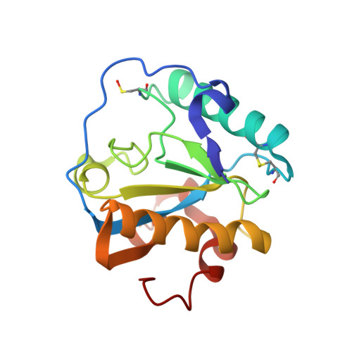
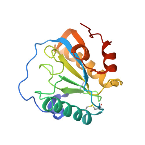
Structure of tracheal cytotoxin in complex with a heterodimeric pattern-recognition receptor.

Chang, C.I.Chelliah, Y.Borek, D.Mengin-Lecreulx, D.Deisenhofer, J.

(2006) Science 311: 1761-1764

  • DOI: https://doi.org/10.1126/science.1123056
  • Primary Citation Related Structures: 
    2F2L

  • PubMed Abstract: 

    Tracheal cytotoxin (TCT), a naturally occurring fragment of Gram-negative peptidoglycan, is a potent elicitor of innate immune responses in Drosophila. It induces the heterodimerization of its recognition receptors, the peptidoglycan recognition proteins (PGRPs) LCa and LCx, which activates the immune deficiency pathway. The crystal structure at 2.1 angstrom resolution of TCT in complex with the ectodomains of PGRP-LCa and PGRP-LCx shows that TCT is bound to and presented by the LCx ectodomain for recognition by the LCa ectodomain; the latter lacks a canonical peptidoglycan-docking groove conserved in other PGRPs. The interface, revealed in atomic detail, between TCT and the receptor complex highlights the importance of the anhydro-containing disaccharide in bridging the two ectodomains together and the critical role of diaminopimelic acid as the specificity determinant for PGRP interaction.


  • Organizational Affiliation
    • Howard Hughes Medical Institute, University of Texas Southwestern Medical Center at Dallas, 6001 Forest Park Road, Dallas, TX 75390-9050, USA.

Macromolecule Content 

  • Total Structure Weight: 39.54 kDa 
  • Atom Count: 2,991 
  • Modeled Residue Count: 333 
  • Deposited Residue Count: 334 
  • Unique protein chains: 2

Macromolecules

Find similar proteins by:|  3D Structure
Entity ID: 1
MoleculeChains  Sequence LengthOrganismDetailsImage
Peptidoglycan-recognition protein-LC isoform LCa167Drosophila melanogasterMutation(s): 1 
UniProt
Find proteins for Q8SXQ7 (Drosophila melanogaster)
Explore Q8SXQ7 
Go to UniProtKB:  Q8SXQ7
Entity Groups
Sequence Clusters30% Identity50% Identity70% Identity90% Identity95% Identity100% Identity
UniProt GroupQ8SXQ7
Glycosylation
Glycosylation Sites: 1Go to GlyGen: Q8SXQ7-1
Sequence Annotations
Expand
Reference Sequence
Find similar proteins by:|  3D Structure
Entity ID: 2
MoleculeChains  Sequence LengthOrganismDetailsImage
Peptidoglycan recognition protein-LC isoform LCxB [auth X]167Drosophila melanogasterMutation(s): 0 
UniProt
Find proteins for Q9GNK5 (Drosophila melanogaster)
Explore Q9GNK5 
Go to UniProtKB:  Q9GNK5
Entity Groups
Sequence Clusters30% Identity50% Identity70% Identity90% Identity95% Identity100% Identity
UniProt GroupQ9GNK5
Sequence Annotations
Expand
Reference Sequence

Small Molecules

Ligands 5 Unique
IDChains Name / Formula / InChI Key2D Diagram3D Interactions
MLD

Query on MLD


Download:Ideal Coordinates CCD File
G [auth X]GLCNAC(BETA1-4)-MURNAC(1,6-ANHYDRO)-L-ALA-GAMMA-D-GLU-MESO-A2PM-D-ALA
C37 H59 N7 O20
UPFMKPIBAIPLHT-RSJSDIDPSA-N
HSQ

Query on HSQ


Download:Ideal Coordinates CCD File
D [auth A]2-acetylamino-2-deoxy-alpha-L-idopyranose
C8 H15 N O6
OVRNDRQMDRJTHS-SLBCVNJHSA-N
NAG

Query on NAG


Download:Ideal Coordinates CCD File
C [auth A]2-acetamido-2-deoxy-beta-D-glucopyranose
C8 H15 N O6
OVRNDRQMDRJTHS-FMDGEEDCSA-N
CIT

Query on CIT


Download:Ideal Coordinates CCD File
H [auth X]CITRIC ACID
C6 H8 O7
KRKNYBCHXYNGOX-UHFFFAOYSA-N
SO4

Query on SO4


Download:Ideal Coordinates CCD File
E [auth A],
F [auth X]
SULFATE ION
O4 S
QAOWNCQODCNURD-UHFFFAOYSA-L
Modified Residues  1 Unique
IDChains TypeFormula2D DiagramParent
CSO
Query on CSO
A
L-PEPTIDE LINKINGC3 H7 N O3 SCYS

Experimental Data & Validation

Experimental Data

  • Method: X-RAY DIFFRACTION
  • Resolution: 2.10 Å
  • R-Value Free:  0.214 (Depositor), 0.259 (DCC) 
  • R-Value Work:  0.166 (Depositor) 
  • R-Value Observed: 0.169 (Depositor) 
Space Group: P 21 21 21
Unit Cell:
Length ( Å )Angle ( ˚ )
a = 41.123α = 90
b = 79.695β = 90
c = 114.389γ = 90
Software Package:
Software NamePurpose
REFMACrefinement
HKL-2000data reduction
SCALEPACKdata scaling
PHASERphasing

Structure Validation

View Full Validation Report


Ligand Structure Quality Assessment 


Entry History 

Deposition Data

Revision History  (Full details and data files)

  • Version 1.0: 2006-04-04
    Type: Initial release
  • Version 1.1: 2008-05-01
    Changes: Version format compliance
  • Version 1.2: 2011-07-13
    Changes: Non-polymer description, Version format compliance
  • Version 1.3: 2011-10-05
    Changes: Derived calculations
  • Version 1.4: 2020-07-29
    Type: Remediation
    Reason: Carbohydrate remediation
    Changes: Advisory, Data collection, Database references, Derived calculations, Structure summary
  • Version 1.5: 2023-08-23
    Changes: Data collection, Database references, Refinement description, Structure summary
  • Version 1.6: 2023-11-15
    Changes: Data collection
  • Version 1.7: 2024-11-20
    Changes: Structure summary