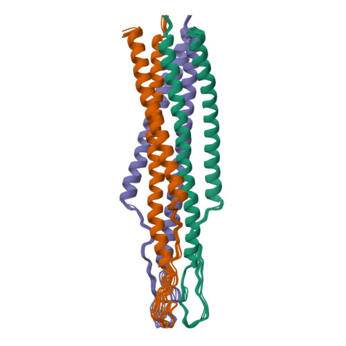
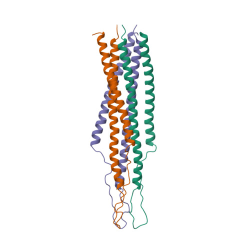
Three-dimensional solution structure of the 44 kDa ectodomain of SIV gp41.
Caffrey, M., Cai, M., Kaufman, J., Stahl, S.J., Wingfield, P.T., Covell, D.G., Gronenborn, A.M., Clore, G.M.(1998) EMBO J 17: 4572-4584
- PubMed: 9707417
- DOI: https://doi.org/10.1093/emboj/17.16.4572
- Primary Citation of Related Structures:
2EZO, 2EZP, 2EZQ, 2EZR, 2EZS - PubMed Abstract:
The solution structure of the ectodomain of simian immunodeficiency virus (SIV) gp41 (e-gp41), consisting of residues 27-149, has been determined by multidimensional heteronuclear NMR spectroscopy. SIV e-gp41 is a symmetric 44 kDa trimer with each subunit consisting of antiparallel N-terminal (residues 30-80) and C-terminal (residues 107-147) helices connected by a 26 residue loop (residues 81-106). The N-terminal helices of each subunit form a parallel coiled-coil structure in the interior of the complex which is surrounded by the C-terminal helices located on the exterior of the complex. The loop region is ordered and displays numerous intermolecular and non-sequential intramolecular contacts. The helical core of SIV e-gp41 is similar to recent X-ray structures of truncated constructs of the helical core of HIV-1 e-gp41. The present structure establishes unambiguously the connectivity of the N- and C-terminal helices in the trimer, and characterizes the conformation of the intervening loop, which has been implicated by mutagenesis and antibody epitope mapping to play a key role in gp120 association. In conjunction with previous studies, the solution structure of the SIV e-gp41 ectodomain provides insight into the binding site of gp120 and the mechanism of cell fusion. The present structure of SIV e-gp41 represents one of the largest protein structures determined by NMR to date.
Organizational Affiliation:
Laboratory of Chemical Physics, Building 5, National Institute of Diabetes and Digestive and Kidney Diseases, National Institutes of Health, Bethesda, MD 20892-0520, USA.


