2EZ0

Crystal structure of the S107A/E148Q/Y445A mutant of EcClC, in complex with a FaB fragment


Experimental Data Snapshot

  • Method: X-RAY DIFFRACTION
  • Resolution: 3.54 Å
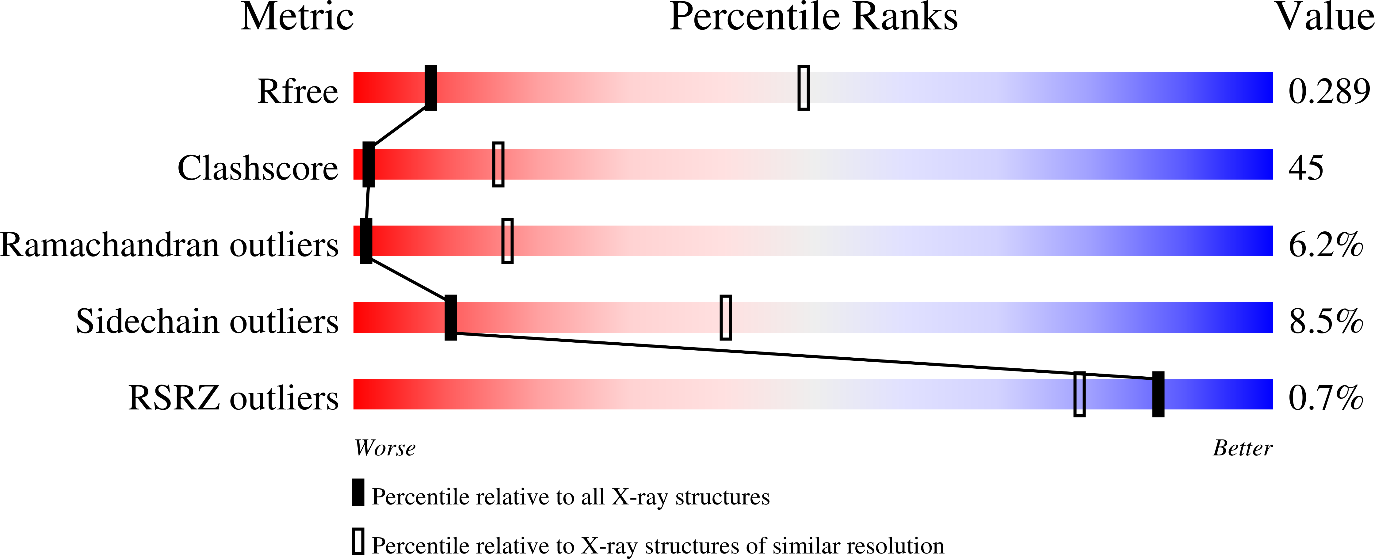
  • R-Value Free: 0.296 
  • R-Value Work: 0.258 
  • R-Value Observed: 0.258 

Starting Model: experimental
View more details

wwPDB Validation   3D Report Full Report


This is version 1.5 of the entry. See complete history


Literature

Ion-binding properties of the ClC chloride selectivity filter.

Lobet, S.Dutzler, R.

(2006) EMBO J 25: 24-33

  • DOI: https://doi.org/10.1038/sj.emboj.7600909
  • Primary Citation of Related Structures:  
    2EXW, 2EXY, 2EZ0

  • PubMed Abstract: 

    The ClC channels are members of a large protein family of chloride (Cl-) channels and secondary active Cl- transporters. Despite their diverse functions, the transmembrane architecture within the family is conserved. Here we present a crystallographic study on the ion-binding properties of the ClC selectivity filter in the close homolog from Escherichia coli (EcClC). The ClC selectivity filter contains three ion-binding sites that bridge the extra- and intracellular solutions. The sites bind Cl- ions with mM affinity. Despite their close proximity within the filter, the three sites can be occupied simultaneously. The ion-binding properties are found conserved from the bacterial transporter EcClC to the human Cl- channel ClC-1, suggesting a close functional link between ion permeation in the channels and active transport in the transporters. In resemblance to K+ channels, ions permeate the ClC channel in a single file, with mutual repulsion between the ions fostering rapid conduction.


  • Organizational Affiliation

    Department of Biochemistry, University of Zürich, Zürich, Switzerland.


Macromolecules
Find similar proteins by:  (by identity cutoff)  |  3D Structure
Entity ID: 1
MoleculeChains Sequence LengthOrganismDetailsImage
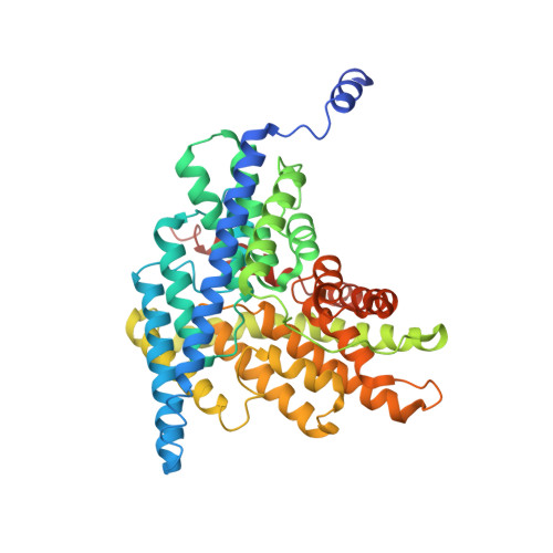
H(+)/Cl(-) exchange transporter clcA
A, B
473Escherichia coliMutation(s): 3 
Membrane Entity: Yes 
UniProt
Find proteins for P37019 (Escherichia coli (strain K12))
Explore P37019 
Go to UniProtKB:  P37019
Entity Groups  
Sequence Clusters30% Identity50% Identity70% Identity90% Identity95% Identity100% Identity
UniProt GroupP37019
Sequence Annotations
Expand
  • Reference Sequence
Find similar proteins by:  (by identity cutoff)  |  3D Structure
Entity ID: 2
MoleculeChains Sequence LengthOrganismDetailsImage
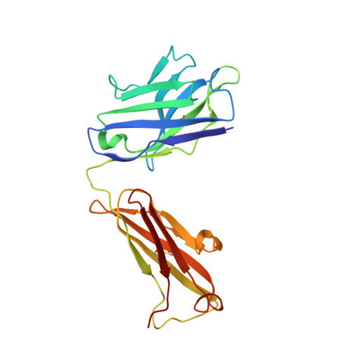
Fab Fragment (Heavy Chain)
C, E
222Mus musculusMutation(s): 0 
Entity Groups  
Sequence Clusters30% Identity50% Identity70% Identity90% Identity95% Identity100% Identity
Sequence Annotations
Expand
  • Reference Sequence
Find similar proteins by:  (by identity cutoff)  |  3D Structure
Entity ID: 3
MoleculeChains Sequence LengthOrganismDetailsImage
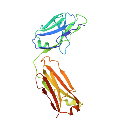
Fab Fragment (Light Chain)
D, F
211Mus musculusMutation(s): 0 
Entity Groups  
Sequence Clusters30% Identity50% Identity70% Identity90% Identity95% Identity100% Identity
Sequence Annotations
Expand
  • Reference Sequence
Experimental Data & Validation

Experimental Data

  • Method: X-RAY DIFFRACTION
  • Resolution: 3.54 Å
  • R-Value Free: 0.296 
  • R-Value Work: 0.258 
  • R-Value Observed: 0.258 
  • Space Group: C 1 2 1
Unit Cell:
Length ( Å )Angle ( ˚ )
a = 220.155α = 90
b = 124.032β = 127.97
c = 149.282γ = 90
Software Package:
Software NamePurpose
CNSrefinement
DENZOdata reduction
SCALEPACKdata scaling
AMoREphasing

Structure Validation

View Full Validation Report


Entry History 

Deposition Data

Revision History  (Full details and data files)

  • Version 1.0: 2006-01-24
    Type: Initial release
  • Version 1.1: 2008-05-01
    Changes: Version format compliance
  • Version 1.2: 2011-07-13
    Changes: Version format compliance
  • Version 1.3: 2021-10-20
    Changes: Database references, Derived calculations
  • Version 1.4: 2023-08-23
    Changes: Data collection, Refinement description
  • Version 1.5: 2024-10-09
    Changes: Structure summary