2EWL | pdb_00002ewl

Solution structure of the C-terminal domain (monomer) of the HPV45 oncoprotein E7


Experimental Data Snapshot

  • Method: SOLUTION NMR
  • Conformers Calculated: 100 
  • Conformers Submitted: 15 
  • Selection Criteria: target function 

wwPDB Validation 3D Report Full Report

Validation slider image for 2EWL

This is version 1.3 of the entry. See complete history

Literature

Solution structure of the partially folded high-risk human papilloma virus 45 oncoprotein E7.

Ohlenschlager, O.Seiboth, T.Zengerling, H.Briese, L.Marchanka, A.Ramachandran, R.Baum, M.Korbas, M.Meyer-Klaucke, W.Durst, M.Gorlach, M.

(2006) Oncogene 25: 5953-5959

  • DOI: https://doi.org/10.1038/sj.onc.1209584
  • Primary Citation Related Structures: 
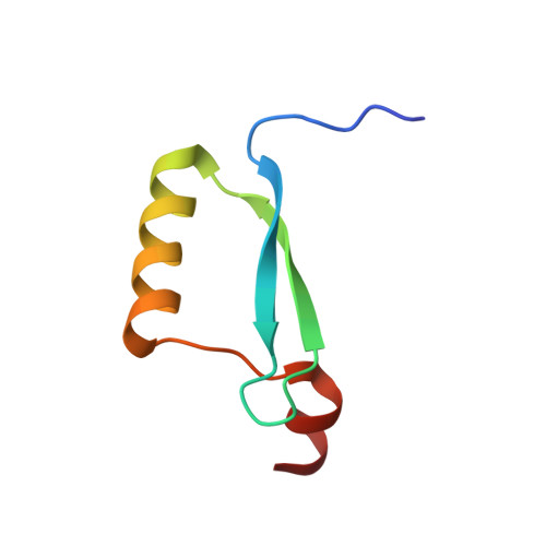
    2EWL, 2F8B

  • PubMed Abstract: 

    The oncoprotein E7 of human papilloma viruses (HPV) is involved in the pathogenesis and maintenance of human cervical cancers. The most prevalent HPV types found in cervix carcinomas are HPV16, 18 and 45. The structure of the E7 dimer from HPV45 (PDB 2F8B) was determined by nuclear magnetic resonance spectroscopy. Each monomer comprises an unfolded N-terminus and a well-structured C-terminal domain with a beta1beta2alpha1beta3alpha2 topology representing a unique zinc-binding fold found only for E7. Dimerization occurs through the alpha1/alpha1' helices and intermolecular beta-sheet formation but excludes the zinc-binding sites. E7 is reported to interact with a number of cellular proteins (e.g. pRb, p21(CIP1)). Binding of a peptide derived from the C-terminus of p21(CIP1) to the C-terminal domain of E7 was characterized by monitoring chemical shift perturbations of the amide groups of E7. This provides direct evidence that a shallow groove situated between alpha1 and beta1 of the E7 C-terminal domain is interacting with the C-terminus of p21(CIP1). Intriguingly, this binding site overlaps with the low-affinity binding site on E7 for the C-domain of pRb.


  • Organizational Affiliation
    • Fritz-Lipmann-Institut, Jena, Germany.

Macromolecule Content 

  • Total Structure Weight: 6.37 kDa 
  • Atom Count: 436 
  • Modeled Residue Count: 56 
  • Deposited Residue Count: 56 
  • Unique protein chains: 1

Macromolecules

Find similar proteins by:|  3D Structure
Entity ID: 1
MoleculeChains  Sequence LengthOrganismDetailsImage
Protein E756human papillomavirus 45Mutation(s): 0 
Gene Names: E7
UniProt
Find proteins for P21736 (Human papillomavirus 45)
Explore P21736 
Go to UniProtKB:  P21736
Entity Groups
Sequence Clusters30% Identity50% Identity70% Identity90% Identity95% Identity100% Identity
UniProt GroupP21736
Sequence Annotations
Expand
Reference Sequence

Small Molecules

Ligands 1 Unique
IDChains Name / Formula / InChI Key2D Diagram3D Interactions
ZN

Query on ZN


Download:Ideal Coordinates CCD File
B [auth A]ZINC ION
Zn
PTFCDOFLOPIGGS-UHFFFAOYSA-N

Experimental Data & Validation

Experimental Data

  • Method: SOLUTION NMR
  • Conformers Calculated: 100 
  • Conformers Submitted: 15 
  • Selection Criteria: target function 

Structure Validation

View Full Validation Report


Entry History 

Deposition Data

Revision History  (Full details and data files)

  • Version 1.0: 2006-10-17
    Type: Initial release
  • Version 1.1: 2008-05-01
    Changes: Version format compliance
  • Version 1.2: 2011-07-13
    Changes: Source and taxonomy, Version format compliance
  • Version 1.3: 2024-05-01
    Changes: Data collection, Database references, Derived calculations