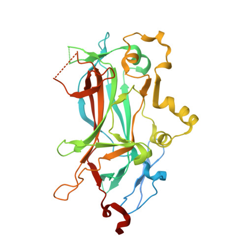
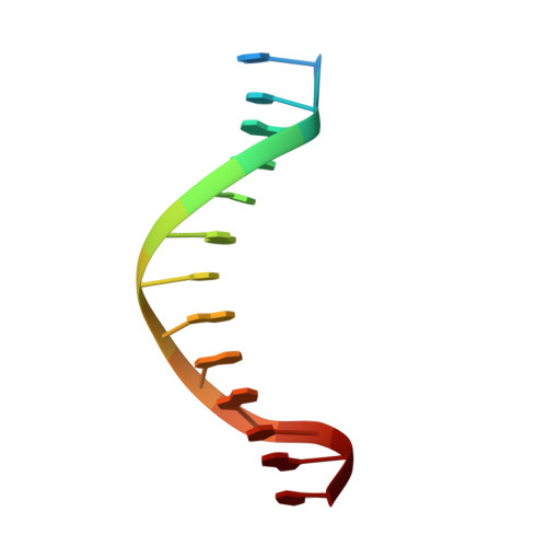
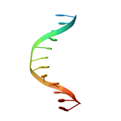
2EUV

Principles of protein-DNA recognition revealed in the structural analysis of Ndt80-MSE DNA complexes


Experimental Data Snapshot

  • Method: X-RAY DIFFRACTION
  • Resolution: 1.94 Å
  • R-Value Free: 0.233 
  • R-Value Work: 0.193 
  • R-Value Observed: 0.195 

Starting Model: experimental
View more details

wwPDB Validation   3D Report Full Report


This is version 1.4 of the entry. See complete history


Literature

Principles of Protein-DNA Recognition Revealed in the Structural Analysis of Ndt80-MSE DNA Complexes.

Lamoureux, J.S.Glover, J.N.

(2006) Structure 14: 555-565

  • DOI: https://doi.org/10.1016/j.str.2005.11.017
  • Primary Citation of Related Structures:  
    2ETW, 2EUV, 2EUW, 2EUX, 2EUZ, 2EVF, 2EVG, 2EVH, 2EVI, 2EVJ

  • PubMed Abstract: 

    The Saccharomyces cerevisiae transcription factor Ndt80 selectively binds a DNA consensus sequence (the middle sporulation element [MSE]) to activate gene expression after the successful completion of meiotic recombination. Here we report the X-ray crystal structures of Ndt80 bound to ten distinct MSE variants. Comparison of these structures with the structure of Ndt80 bound to a consensus MSE reveals structural principles that determine the DNA binding specificity of this transcription factor. The 5' GC-rich end of the MSE contains distinct 5'-YpG-3' steps that are recognized by arginine side chains through a combination of hydrogen bonding and cation-pi interactions. The 3' AT-rich region is recognized via minor groove contacts that sterically exclude the N2 atom of GC base pairs. The conformation of the AT-rich region is fixed by interactions with the protein that favor recognition of poly(A)-poly(T) versus mixed AT sequences through an avoidance of major groove steric clashes at 5'-ApT-3' steps.


  • Organizational Affiliation

    Department of Biochemistry, University of Alberta, Edmonton, Alberta T6G 2H7, Canada.


Macromolecules

Find similar proteins by:  (by identity cutoff)  |  3D Structure
Entity ID: 3
MoleculeChains Sequence LengthOrganismDetailsImage
NDT80 proteinC [auth A]345Saccharomyces cerevisiaeMutation(s): 2 
Gene Names: ndt80
UniProt
Find proteins for P38830 (Saccharomyces cerevisiae (strain ATCC 204508 / S288c))
Explore P38830 
Go to UniProtKB:  P38830
Entity Groups  
Sequence Clusters30% Identity50% Identity70% Identity90% Identity95% Identity100% Identity
UniProt GroupP38830
Sequence Annotations
Expand
  • Reference Sequence

Find similar nucleic acids by:  Sequence   |   3D Structure  

Entity ID: 1
MoleculeChains LengthOrganismImage
5'-D(*TP*GP*CP*AP*AP*CP*AP*CP*AP*AP*AP*TP*AP*C)-3'A [auth B]14N/A
Sequence Annotations
Expand
  • Reference Sequence

Find similar nucleic acids by:  Sequence   |   3D Structure  

Entity ID: 2
MoleculeChains LengthOrganismImage
5'-D(*AP*GP*TP*AP*TP*TP*TP*GP*TP*GP*TP*TP*GP*C)-3'B [auth C]14N/A
Sequence Annotations
Expand
  • Reference Sequence
Experimental Data & Validation

Experimental Data

  • Method: X-RAY DIFFRACTION
  • Resolution: 1.94 Å
  • R-Value Free: 0.233 
  • R-Value Work: 0.193 
  • R-Value Observed: 0.195 
  • Space Group: C 2 2 21
Unit Cell:
Length ( Å )Angle ( ˚ )
a = 69.226α = 90
b = 79.248β = 90
c = 160.881γ = 90
Software Package:
Software NamePurpose
SCALAdata scaling
REFMACrefinement
PDB_EXTRACTdata extraction
MOSFLMdata reduction
CCP4data scaling

Structure Validation

View Full Validation Report


Entry History 

Deposition Data

Revision History  (Full details and data files)

  • Version 1.0: 2006-03-21
    Type: Initial release
  • Version 1.1: 2008-05-01
    Changes: Version format compliance
  • Version 1.2: 2011-07-13
    Changes: Version format compliance
  • Version 1.3: 2021-10-20
    Changes: Database references, Derived calculations
  • Version 1.4: 2023-08-23
    Changes: Data collection, Refinement description