2ER0 | pdb_00002er0

X-RAY STUDIES OF ASPARTIC PROTEINASE-STATINE INHIBITOR COMPLEXES


Experimental Data Snapshot

  • Method: X-RAY DIFFRACTION
  • Resolution: 3.00 Å
  • R-Value Work: 
    0.280 (Depositor) 

wwPDB Validation 3D Report Full Report

Validation slider image for 2ER0

This is version 1.4 of the entry. See complete history

Literature

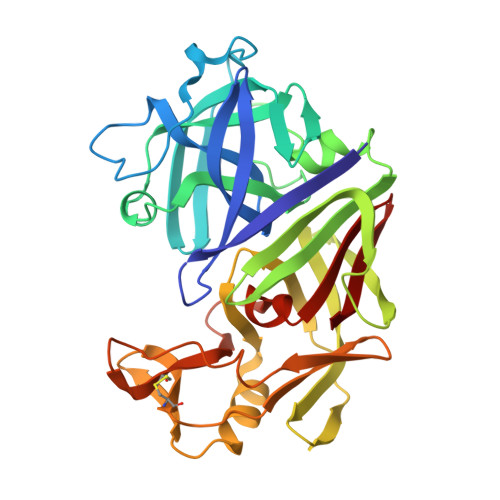
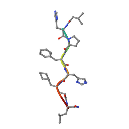
X-ray studies of aspartic proteinase-statine inhibitor complexes.

Cooper, J.B.Foundling, S.I.Blundell, T.L.Boger, J.Jupp, R.A.Kay, J.

(1989) Biochemistry 28: 8596-8603

  • DOI: https://doi.org/10.1021/bi00447a049
  • Primary Citation Related Structures: 
    2ER0, 2ER9

  • PubMed Abstract: 

    The conformation of a statine-containing renin inhibitor complexed with the aspartic proteinase from the fungus Endothia parasitica (EC 3.4.23.6) has been determined by X-ray diffraction at 2.2-A resolution (R = 0.17). We describe the structure of the complex at high resolution and compare this with a 3.0-A resolution analysis of a bound inhibitor, L-364,099, containing a cyclohexylalanine analogue of statine. The inhibitors bind in extended conformations in the long active-site cleft, and the hydroxyl of the transition-state analogue, statine, interacts strongly with the catalytic aspartates via hydrogen bonds to the essential carboxyl groups. This work provides a detailed structural analysis of the role of statine in peptide inhibitors. It shows conclusively that statine should be considered a dipeptide analogue (occupying P1 to P1') despite lacking the equivalent of a P1' side chain, although other inhibitor residues (especially P2) may compensate by interacting at the unoccupied S1' specificity subsite.


  • Organizational Affiliation
    • Department of Crystallography, Birkbeck College, London, U.K.

Macromolecule Content 

  • Total Structure Weight: 34.89 kDa 
  • Atom Count: 2,456 
  • Modeled Residue Count: 338 
  • Deposited Residue Count: 338 
  • Unique protein chains: 2

Macromolecules

Find similar proteins by:|  3D Structure
Entity ID: 1
MoleculeChains  Sequence LengthOrganismDetailsImage
ENDOTHIAPEPSINA [auth E]330Cryphonectria parasiticaMutation(s): 0 
EC: 3.4.23.6 (PDB Primary Data), 3.4.23.22 (UniProt)
UniProt
Find proteins for P11838 (Cryphonectria parasitica)
Explore P11838 
Go to UniProtKB:  P11838
Entity Groups
Sequence Clusters30% Identity50% Identity70% Identity90% Identity95% Identity100% Identity
UniProt GroupP11838
Sequence Annotations
Expand
Reference Sequence
Find similar proteins by:  Sequence   |   3D Structure  
Entity ID: 2
MoleculeChains  Sequence LengthOrganismDetailsImage
L364,099B [auth I]8N/AMutation(s): 0 

Experimental Data & Validation

Experimental Data

  • Method: X-RAY DIFFRACTION
  • Resolution: 3.00 Å
  • R-Value Work:  0.280 (Depositor) 
Space Group: P 1 21 1
Unit Cell:
Length ( Å )Angle ( ˚ )
a = 43α = 90
b = 75.8β = 97
c = 42.8γ = 90
Software Package:
Software NamePurpose
PROLSQrefinement

Structure Validation

View Full Validation Report


Entry History 

Deposition Data

Revision History  (Full details and data files)

  • Version 1.0: 1991-01-15
    Type: Initial release
  • Version 1.1: 2008-03-03
    Changes: Version format compliance
  • Version 1.2: 2011-07-13
    Changes: Atomic model, Database references, Derived calculations, Non-polymer description, Structure summary, Version format compliance
  • Version 1.3: 2017-11-29
    Changes: Derived calculations, Other
  • Version 1.4: 2024-11-06
    Changes: Data collection, Database references, Derived calculations, Structure summary