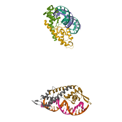
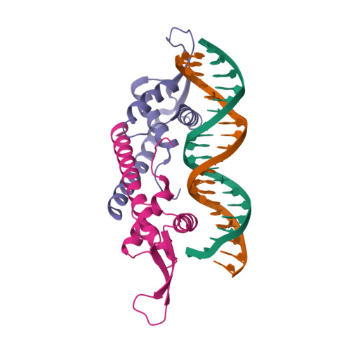
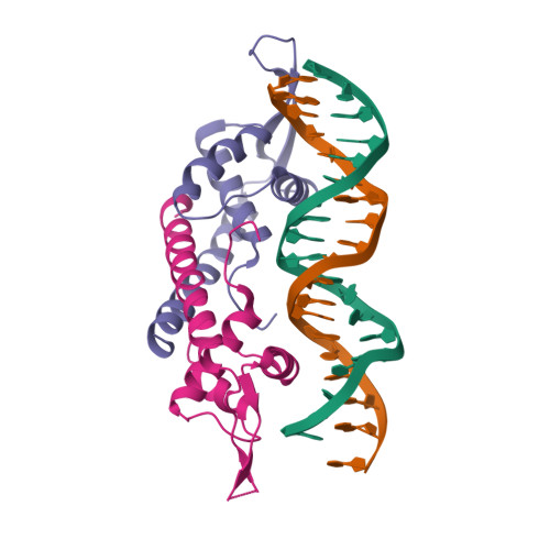
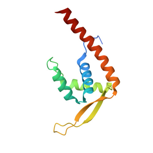
2EFW

Crystal structure of the RTP:nRB complex from Bacillus subtilis


Experimental Data Snapshot

  • Method: X-RAY DIFFRACTION
  • Resolution: 2.50 Å
  • R-Value Free: 0.332 
  • R-Value Work: 0.291 
  • R-Value Observed: 0.293 

Starting Model: experimental
View more details

wwPDB Validation   3D Report Full Report


This is version 1.3 of the entry. See complete history


Literature

An asymmetric structure of the Bacillus subtilis replication terminator protein in complex with DNA

Vivian, J.P.Porter, C.J.Wilce, J.A.Wilce, M.C.J.

(2007) J Mol Biol 370: 481-491

  • DOI: https://doi.org/10.1016/j.jmb.2007.02.067
  • Primary Citation of Related Structures:  
    2EFW

  • PubMed Abstract: 

    In Bacillus subtilis, the termination of DNA replication via polar fork arrest is effected by a specific protein:DNA complex formed between the replication terminator protein (RTP) and DNA terminator sites. We report the crystal structure of a replication terminator protein homologue (RTP.C110S) of B. subtilis in complex with the high affinity component of one of its cognate DNA termination sites, known as the TerI B-site, refined at 2.5 A resolution. The 21 bp RTP:DNA complex displays marked structural asymmetry in both the homodimeric protein and the DNA. This is in contrast to the previously reported complex formed with a symmetrical TerI B-site homologue. The induced asymmetry is consistent with the complex's solution properties as determined using NMR spectroscopy. Concomitant with this asymmetry is variation in the protein:DNA binding pattern for each of the subunits of the RTP homodimer. It is proposed that the asymmetric "wing" positions, as well as other asymmetrical features of the RTP:DNA complex, are critical for the cooperative binding that underlies the mechanism of polar fork arrest at the complete terminator site.


  • Organizational Affiliation

    Department of Pharmacology, University of Western Australia, Nedlands, Western Australia, 6009, Australia.


Macromolecules

Find similar proteins by:  (by identity cutoff)  |  3D Structure
Entity ID: 3
MoleculeChains Sequence LengthOrganismDetailsImage
Replication termination proteinE [auth A],
F [auth B],
G [auth F],
H [auth G]
122Bacillus subtilisMutation(s): 1 
Gene Names: rtp
UniProt
Find proteins for P0CI76 (Bacillus subtilis (strain 168))
Explore P0CI76 
Go to UniProtKB:  P0CI76
Entity Groups  
Sequence Clusters30% Identity50% Identity70% Identity90% Identity95% Identity100% Identity
UniProt GroupP0CI76
Sequence Annotations
Expand
  • Reference Sequence

Find similar nucleic acids by:  Sequence   |   3D Structure  

Entity ID: 1
MoleculeChains LengthOrganismImage
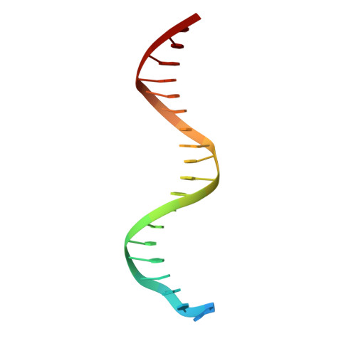
DNA (5'-D(*DCP*DT*DAP*DTP*DGP*DTP*DAP*DCP*DCP*DAP*DAP*DAP*DTP*DGP*DTP*DTP*DCP*DAP*DGP*DTP*DC)-3')A [auth D],
C [auth I]
21N/A
Sequence Annotations
Expand
  • Reference Sequence

Find similar nucleic acids by:  Sequence   |   3D Structure  

Entity ID: 2
MoleculeChains LengthOrganismImage
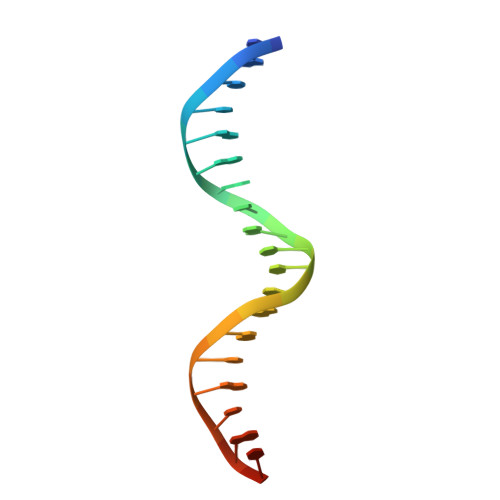
DNA (5'-D(*DGP*DAP*DCP*DTP*DGP*DAP*DAP*DCP*DAP*DTP*DTP*DTP*DGP*DGP*DTP*DAP*DCP*DAP*DTP*DAP*DG)-3')B [auth E],
D [auth J]
21N/A
Sequence Annotations
Expand
  • Reference Sequence
Experimental Data & Validation

Experimental Data

  • Method: X-RAY DIFFRACTION
  • Resolution: 2.50 Å
  • R-Value Free: 0.332 
  • R-Value Work: 0.291 
  • R-Value Observed: 0.293 
  • Space Group: C 2 2 21
Unit Cell:
Length ( Å )Angle ( ˚ )
a = 124.012α = 90
b = 129.626β = 90
c = 125.221γ = 90
Software Package:
Software NamePurpose
REFMACrefinement
ADSCdata collection
DENZOdata reduction
SCALEPACKdata scaling
MOLREPphasing

Structure Validation

View Full Validation Report


Entry History 

Deposition Data

Revision History  (Full details and data files)

  • Version 1.0: 2008-02-26
    Type: Initial release
  • Version 1.1: 2011-07-13
    Changes: Advisory, Version format compliance
  • Version 1.2: 2021-11-10
    Changes: Database references
  • Version 1.3: 2023-10-25
    Changes: Data collection, Refinement description